Endodontic Management and Adhesive Rehabilitation of Grossly Decayed Mandibular First Molar Using Lithium Disilicate Endocrown: A Clinical Case Report
Department of Conservative Dentistry and Endodontics, Seema Dental College And Hospital, Rishikesh
ABSTRACT
Management of severely mutilated posterior teeth requires both successful endodontic therapy and adequate post-endodontic rehabilitation. Endocrowns have emerged as a conservative restorative alternative that eliminates the need for intra-radicular posts. This case report describes nonsurgical root canal treatment followed by CAD-CAM fabricated lithium disilicate endocrown restoration of grossly decayed mandibular first molars. Clinical outcomes were satisfactory, demonstrating functional and biomechanical success.
KEYWORDS: Root canal treatment, Endocrown, Lithium disilicate, Adhesive cementation, CAD-CAM
INTRODUCTION
Endodontically treated teeth are structurally compromised due to loss of tooth structure and altered biomechanical properties. Conventional post-core supported crowns may increase the risk of root fracture due to removal of radicular dentin. The endocrown concept utilizes the pulp chamber for retention and adhesive bonding for stabilization, thereby preserving tooth structure and improving stress distribution. This report presents combined endodontic and restorative management of severely damaged mandibular molars.
CASE REPORT
Case 1
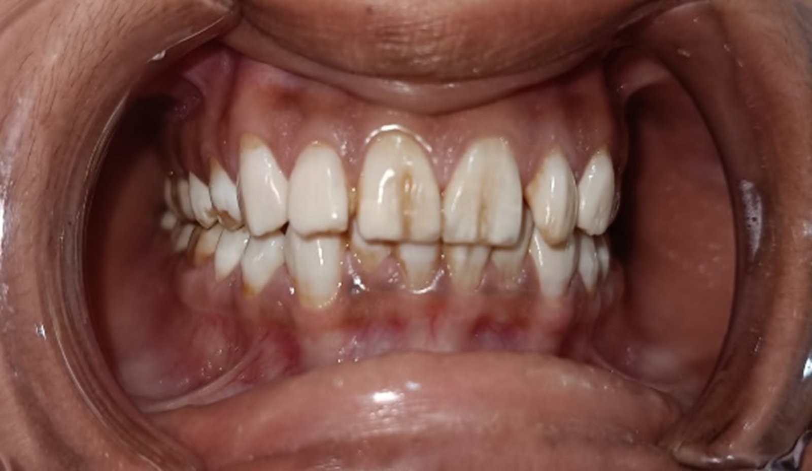
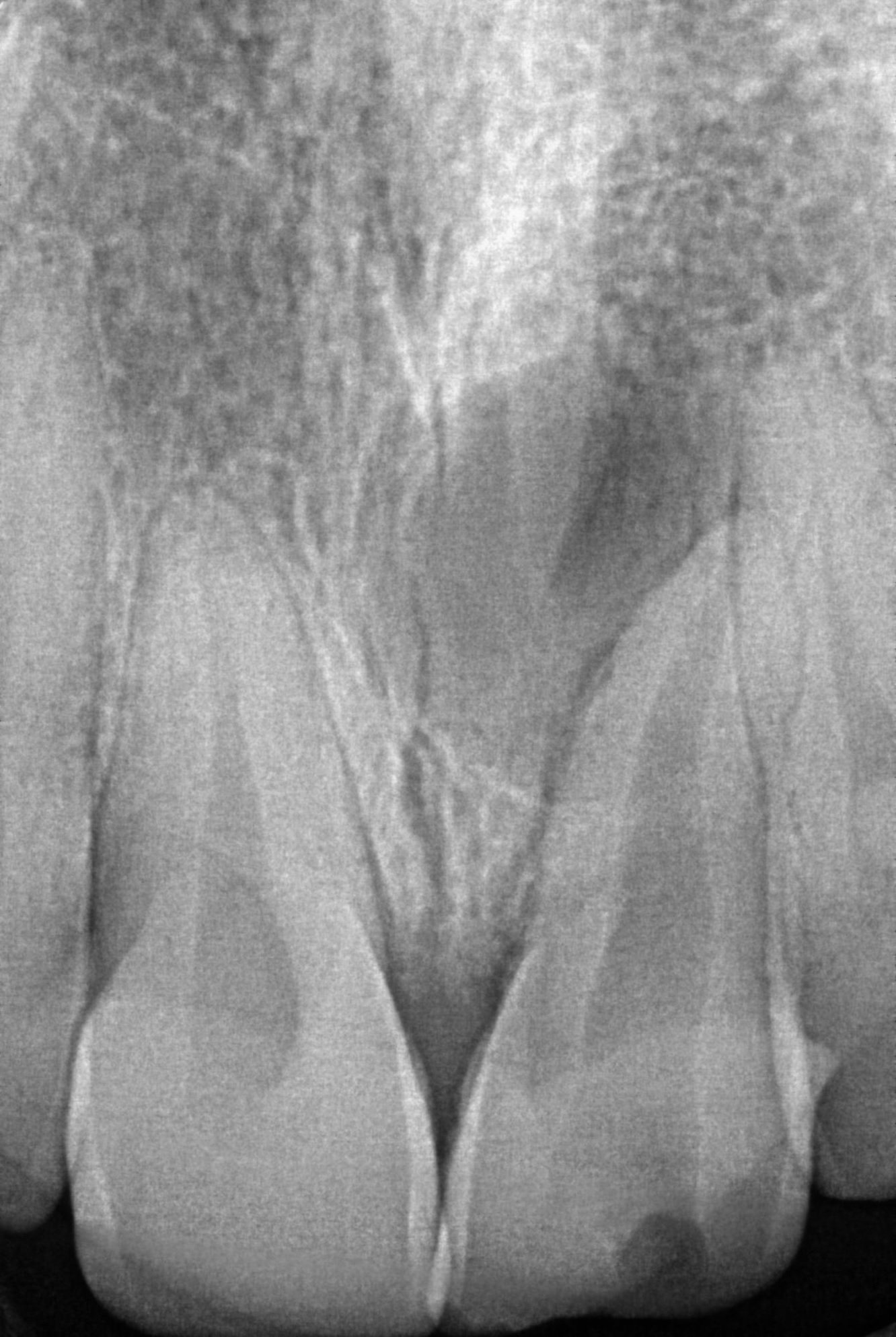
A 30-year-old patient reported with pain and food lodgment in the lower left posterior region. Clinical examination revealed a deep carious lesion with extensive coronal destruction in relation to the mandibular left first molar. The tooth was tender on percussion. Based on clinical findings, a diagnosis of symptomatic irreversible pulpitis with apical involvement was made.
Endodontic Procedure
After administration of local anesthesia and rubber dam isolation, access cavity preparation was performed. Working length was determined using an electronic apex locator. Biomechanical preparation was carried out using rotary nickel-titanium instruments with copious irrigation using sodium hypochlorite and saline. Final irrigation included EDTA for smear layer removal.
Obturation was performed using gutta-percha cones and resin-based sealer by lateral condensation technique.
Post-Endodontic Restoration
Due to extensive coronal destruction and adequate pulp chamber depth, endocrown restoration was planned.
Endocrown Restorative Procedure
Tooth preparation was done to obtain a circumferential butt-joint margin of approximately 1–1.5 mm with occlusal reduction of about 2 mm. The pulp chamber was prepared to provide internal retention.
Digital impression was recorded using an intraoral scanner. The restoration was designed and milled from lithium disilicate ceramic using CAD-CAM technology.
Try-in was performed to evaluate marginal adaptation, proximal contacts, and occlusion. Adhesive cementation was carried out using dual-cure resin cement. Finishing and polishing were completed.


Case 2
A 26-year-old patient reported with pain and food lodgment in the lower right posterior region. Clinical examination revealed a deep carious lesion with extensive coronal destruction in relation to the mandibular right first molar.
The treatment protocol followed was similar to Case 1, including root canal treatment and rehabilitation with lithium disilicate endocrown.


RESULT AND FOLLOW-UP
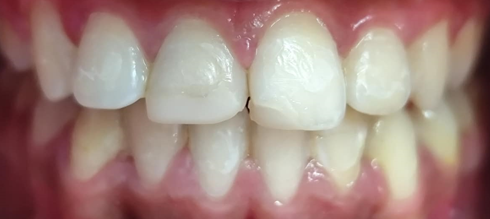
The restorations were evaluated clinically for marginal adaptation, proximal contacts, and occlusion. Both endocrowns demonstrated satisfactory fit and stability.
At follow-up visits, the treated teeth remained asymptomatic with no signs of pain, tenderness, or functional discomfort. Patients reported improved mastication and comfort. Surrounding periodontal tissues were healthy, indicating successful clinical outcomes.
DISCUSSION
The prognosis of endodontically treated posterior teeth largely depends on effective coronal rehabilitation and preservation of remaining tooth structure. Conventional post-core restorations may weaken radicular dentin and increase fracture risk. Endocrowns provide a conservative alternative by utilizing the pulp chamber for retention and adhesive bonding to form a biomechanically stable monoblock restoration.
In these cases, adequate cervical enamel and pulp chamber depth supported the use of a butt-joint preparation design, promoting favorable stress distribution. The use of lithium disilicate ceramic combined with CAD-CAM fabrication and adhesive cementation contributed to satisfactory marginal adaptation, functional performance, and esthetic integration.
CONCLUSION
Endocrown restoration following endodontic therapy represents an effective and minimally invasive approach for rehabilitation of grossly decayed mandibular molars, providing satisfactory clinical performance.
REFERENCES
- Bindl A, Mörmann WH. Clinical evaluation of adhesively placed ceramic endocrowns after 2 years. Int J Prosthodont. 1999;12:395–402.
- Belleflamme MM, Geerts SO, Louwette MM, Grenade CF, Vanheusden AJ, Mainjot AK. No post–no core approach to restore severely damaged posterior teeth: Up to 10-year retrospective study. J Dent. 2017;63:1–7.
- Rocca GT, Krejci I. Crown and post-free adhesive restorations for endodontically treated posterior teeth: From direct composite to endocrowns. Eur J Esthet Dent. 2013;8:156–179.
- Fages M, Bennasar B. The endocrown: A different type of all-ceramic reconstruction for molars. J Can Dent Assoc. 2013;79:d140.
- Biacchi GR, Basting RT. Comparison of fracture strength of endocrowns and glass fiber post-retained conventional crowns. Oper Dent. 2012;37:130–136.
- Pissis P. Fabrication of a metal-free ceramic restoration utilizing the monobloc technique. Pract Periodontics Aesthet Dent. 1995;7:83–94.
- Ingle JI, Bakland LK, Baumgartner JC. Ingle’s Endodontics. 6th ed. Hamilton: BC Decker; 2008.
Disclaimer from Guident
The views and opinions expressed in articles published in Guident are solely those of the authors and do not necessarily reflect the views of the editors, the editorial board or the publisher, Ivory India. Guident does not endorse any product, technique or treatment described. Clinical content is intended for qualified dental and medical professionals and is not a substitute for individual clinical judgement. Guident, its editors and the publisher accept no liability for any consequence arising from the use of information published in the journal.
How to cite this article
Dr. Deepshikha Sainwal, Dr. Vidya Chawla, Dr. Seema Dixit. Endodontic Management and Adhesive Rehabilitation of Grossly Decayed Mandibular First Molar Using Lithium Disilicate Endocrown: A Clinical Case Report. Guident. 2026 Jun;19(7). Available from: https://ivoryindia.in/articles/endodontics/endodontic-management-and-adhesive-rehabilitation-of-grossly-decayed-mandibular-first-molar-using-lithium.html