3D Printed Bite Registration Jig BRJ - a Key to Distal Extension Implant Cases
Department of Prosthodontics & Implantology K.D. Dental College & Hospital, Mathura (U.P) India
INTRODUCTION
Clinicians often face challenges while working with patients who are completely edentulous or with a failing dentition and request treatment. Rehabilitation of the dentition with removable or fixed restorations with dental implants remains an exciting, yet challenging, dental procedure.
Fixed rehabilitation with dental implants requires precise surgical as well as prosthetic techniques that enable a clinician to deliver a prosthesis accomplishing patient’s aesthetic, functional, and maintenance goals. Fixed restorations are often technically demanding in regard to implant position, angulation, tooth position, emergence form, and the interplay between teeth and tissues. Additionally, other factors related to patient-mediated outcomes, occlusal scheme, implant number, and implant position within the arch further complicate planning rehabilitation with fixed prostheses.
The first step of successful restorative procedures is the accurate transfer of intra-oral information for extraoral use. Moreover, appropriate articulation is necessary to transfer this intra-oral information to a maxillary and a mandibular cast which require establishment of three points of contact. These points of contact should be adequately spaced so as to provide adequate stability. Missing teeth eliminates the more traditional means of stabilization. If the missing teeth are distally located, the soft tissue of the edentulous region has to be utilized to establish the tripod. Therefore, an adequate bite registration is the key to provide the pertinent orientation between the maxillary and mandibular casts but is often overlooked.
3D PRINTING
3D printing also referred to as “additive manufacturing” is a method which uses 3 dimensional CAD data sets for producing 3D physical models. It is the method of fabricating material of choice namely, metal/polymer/resins, in incremental layers using CAD/CAM (Computer Aided Design/Computer Aided Manufacturing) digital scans for the dual purpose of designing a product and for controlling the manufacturing process.
The procedure involves giving input to the computer through 3D CAD data which materializes the design by additive deposition of the material of choice to give final shape to the desired model. The first ever use of 3D printing in dentistry was in 1999.
High accuracy can be achieved through 3D printed templates for guided implant surgery when used as alternatives to laboratory manufactured templates. The guides offer better precision in placement of implant. Also, the cost effectiveness and time saving fabrication of the guides adds an advantage in to 3D data analysis and interpretation which ensures more predictable results.
3D BITE REGISTRATION JIG V/S OTHER TECHNIQUES
Techniques of bite registration for distal extension implant restorations includes the conventional - occlusal rim, temporary cylinders, auto polymerizing resin - healing abutments, bite registration aid and BRJ.
Conventional method incorporates construction of a base and a wax occlusal rim to provide appropriate support for the registration material and to facilitate mounting of an opposing cast (Figure 1). The extra time is required to fabricate the cast and record base which increases cost and appointments. Inaccuracies due to compression of the ridge under the record base may also be incorporated.
Temporary cylinders are used by engaging them on a distal implant at the time of the fixture level impression (Figure 2). Bite registration material is then injected around the cylinder to record the bite when in maximum intercuspation, eliminating an additional patient visit. The disadvantages with temporary cylinders are that they are expensive, non-reusable and uncomfortable for the patient when adjusted in the mouth. Also, they might not be long enough which necessitate the use of excessive bite registration material leading to discrepancies such as shrinkage (auto polymerizing acrylic resin).
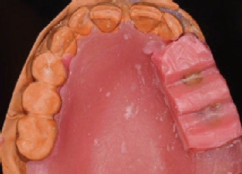
Figure 1: Conventional interocclusal registration technique utilizing record base and occlusal rim

Figure 2: Interocclusal registration technique utilizing temporary cylinder and autopolymerizing acrylic resin.
Healing abutments located within an edentulous distal extension space can be used as an alternative technique (Figure 3). They act as vertical stops for an interocclusal record with the use of acrylic resin. This technique requires positioning the same healing abutments that the patient has in the mouth on the cast, which thereby, increases the fee and patient discomfort due to the acrylic resin. Also, the position of healing abutment intraorally will not exactly be the same on cast due to the different torque and the nonengaging nature of the healing abutments. Another disadvantage is that the healing abutments are short, so the space between them and the opposing dentition is usually more than 5mm resulting in the use of undesirable amount of autopolymerizing acrylic resin to fill the space leading to shrinkage and possible inaccuracy.
In contrast to other techniques, one manufacturer has developed a specific plastic “bite registration aid” (Straumann AG, Basel, Switzerland), which can be specifically used on their dental implants in order to obtain and transfer an interocclusal record (Figure 4). This bite registration aid though obliterates the construction of a tissue supported record base, it still remains expensive and is disposable. Moreover, its use is limited to one implant system.

Figure 3: Interocclusal registration technique utilizing healing abutment and autopolymerizing acrylic resin.

Figure 4: Bite registration aid.
Recent technique for bite registration is the use of 3D printed BRJ. This BRJ have retention grooves fabricated by the printer (Figure 5 and 6). The clinical procedure includes: Pick up impression using a polyether material. At the same appointment, bite registration is performed by engaging one BRJ on the distal implant (Figures 7). Vertical height is then adjusted, using a round - end tapered carbide bur, intraorally. Bite registration material is injected around the BRJ and the patient is instructed to close in a centric relation position. Subsequently, the BRJ, embedded in the bite registration material, is used extraorally to mount the fixture level cast with the opposing cast (Figures 8 and 9). Finally, insertion of the prosthesis at the try-in procedure is done.

Figure 5: 3D printed BRJ.

Figure 6: BRJ with implant analogue.

Figure 7: BRJ engaged on implant analogue.

Figure 8: The BRJ embedded with bite registration material as the interocclusal record.

Figure 9: Articulation of fixture cast and opposing cast utilizing the BRJ interocclusal record.
DISCUSSION
A comparison of similar techniques of bite registration for distal extension implant restorations is given in Table 1.
Table 1: Comparison of various bite registration techniques for distal extension implant restorations.

CONCLUSION
3D printing due to its efficacy, accuracy, potency and less time consumption in the fabrication process could establish itself as a milestone in the field of dentistry. Use of 3D printing in treatment planning and analysis of treatment outcomes improves the quality of treatment enhancing patient satisfaction. BRJ provides a practical and time saving method by recording the bite at the time of the impression making thus, reducing patient visits. Moreover, it provides more precision as compared to other techniques for bite registration for distal extension implant cases, due to its anti-rotational feature, narrow interface (platform switch design), no interference with the soft tissue and adequate height which reduces the amount of bite registration material that might cause discrepancies. Another benefit with BRJs is that it can be used with any implant system. In addition, 3-D printing of one hundred BRJs takes around two hours and less than one dollar which makes this technique feasible and beneficial.
REFERENCES
- Lagoudis M, Ceballos L, Manasse M, Trushkowsky RD, et al. 3-D Printed Bite Registration Jigs for Distal Free End Implant Restorations. J Oral Bio. 2016; 3(2): 4.
- Rengier F, Mehndiratta A, von Tengg-Kobligk H et al. 3D printing based on imaging data: Review of medical applications. Int J CARS. 2010; 5(4):335–41. crossref PMid:20467825
- Dawood A, Marti B, Jackson VS, et al. 3D printing in dentistry. British Dental Journal. 2015; 219:521–9. crossref PMid:26657435
- Perakis N, Belser UC, Magne P (2004) Final impressions: a review of material properties and description of a current technique. Int J Periodontics Restorative Dent 24: 109-117.
- Freilich MA, Altieri JV, Wahle JJ (1992) Principles for selecting interocclusal records for articulation of dentate and partially dentate casts. J Prosthet Dent 68: 361-367.
- Goldstein RE (2008) A precise bite registration technique. Inside Dentistry 4.
- Langer A (1981) The validity of maxillomandibular records made with trial and processed acrylic resin bases. J Prosthet Dent 45: 253-258.
- Rosenstiel SF, Land MF, Fujimoto Jc (2006) Contemporary fixed prosthodontics. St. Louis, Mosby, pp. 62.
- Henderson D, Steffel VL (1977) Mc Craken’s removable partial prosthodntics. St. Louis, Mosby, pp. 290-291.
- Wilhelm MW (2013) New method for improving the accuracy of bite registrations in restorative implant dentistry. Compendium 34.
- Wicks R, Ahuja S, Jain V, Ferreira CF (2013) A technique to create an interocclusal bite registration using in situ implant healing abutments. J Tenn Dent Assoc 93: 47-49.
Disclaimer from Guident
The views and opinions expressed in articles published in Guident are solely those of the authors and do not necessarily reflect the views of the editors, the editorial board or the publisher, Ivory India. Guident does not endorse any product, technique or treatment described. Clinical content is intended for qualified dental and medical professionals and is not a substitute for individual clinical judgement. Guident, its editors and the publisher accept no liability for any consequence arising from the use of information published in the journal.
How to cite this article
Dr. Yashi Garg, Dr. Adil Khursheed Sheikh, Dr. Manesh Lahori. 3D Printed Bite Registration Jig BRJ - a Key to Distal Extension Implant Cases. Guident. 2019 Sep;12(10). Available from: https://ivoryindia.in/articles/implantology/3d-printed-bite-registration-jig-brj-a-key-to-distal-extension-implant-cases.html