Immediate Temporization in the Aesthetic Zone Using Noble Active Ti Ultra Implant: A Case Report
Wisdom Dental Clinics ®, Jaipur, India
ABSTRACT
The aesthetic and functional integrity of the periodontal tissues may be compromised by dental loss. Dental implants have become an additional tool in armamentarium of treatment options to offer patient for replacement of missing tooth or teeth. Diagnosis and treatment planning is key factors in achieving the successful outcome after placing and restoring implants placed immediately after tooth extraction. This case report demonstrates the use of immediate implant placement in anterior maxillary region followed by temporization and permanent crown placement after one and half month.
Keywords: immediate implant, aesthetic, anterior region
INTRODUCTION
Dental Implants are today considered as a reliable treatment option to replace missing teeth both for aesthetics and function.1 Earlier in 70s and 80s implants were placed in healed alveolar ridges.2 It was in the 90s that implant placement started in fresh extraction sockets or in partially healed alveolar ridges predominantly for implants in the aesthetic zone.3,4 Implant placement in anterior maxilla area is challenging because of aesthetic concern and unfavourable bone topography. The success of an implant restoration depends on proper implant placement and the hard and soft tissue architecture that surrounds the fixture.5
According to the first well-established protocol for the modern dental implants, implants were surgically placed in the jaws only after a 3–6-month period after tooth extraction, to ensure satisfactory remodeling and healing of the alveolar bone in order to optimize osseointegration of the implant.6 Nowadays, it has been widely accepted that dental implants can be inserted into the alveolar socket immediately after extraction of teeth, with survival rates comparable to the ones observed for implants placed in pristine or healed sites.7,8
The immediate implant placement (IIP) approach has its particularities depending on which region of the jaws is involved. The alveolar bone of the anterior maxilla is usually proclined in an anterior-inferior direction, forming a concavity right above the apical region of the teeth, and the labial bone is this region is usually thin.9 Therefore, it is expected that the frequency of bone dehiscence and esthetic problems would be higher in this region in comparison to the posterior regions of the jaw10, as the alveolar ridge is followed by a great reduction of its dimensions after the extraction of a tooth.11 The issue is especially important if the implants planned to be placed immediately after extraction are positioned in the same position as the tooth that previously occupied the socket,12 the so-called prosthetically-driven position, as it is advocated that the implant should be anchored by a minimum amount of bone apical to the socket in order to achieve primary stability.13 Having that in mind, it is important that an adequate pre-treatment evaluation is conducted in the cases for which IIP is planned.14
The placement of a provisional restoration on a single implant in the aesthetic zone has been advocated for creating a good soft tissue contour, especially in conjunction with immediate implant placement.15 Immediate implant placement and provisionalization can be a predictable treatment modality to replace a hopeless tooth in the aesthetic zone when primary implant stability can be achieved, and the provisional restoration can be adjusted to clear all centric and eccentric contacts. This case report discussed the key concepts of treatment planning, implant surgery, and prosthetic rehabilitation needed to achieve aesthetic success in the maxillary anterior region. The use of dental implants in the maxillary anterior region to replace missing teeth is a viable treatment option.
CASE REPORT
A 27-year-old female patient in excellent health reported with sensitivity in maxillary anterior jaw region 21 which has metal ceramic crown placed 10 years ago. [Figure 1] The patient’s medical history was unremarkable and on dental hard tissue examination there were no other significant findings. Overall periodontal status- oral hygiene was good but food lodgement was present in area of crown. Marginal gingival inflammation was detected. No periodontal pockets were present, no other tooth mobility found. The patient desired fixed type replacement which preserved the neighbouring healthy teeth.

Figure 1: Pre-operative photographs
TREATMENT PLANNING
After the clinical examination and review of the initial periapical radiograph, the treatment was planned to remove the metal ceramic crown w.r.t 11 ,21 and place implant w.r.t 21.
The patient was advised of the potential benefits of a CBCT scan, this information would yield the information necessary to recommend the optimal treatment plan, based on a comprehensive assessment of the bone for the potential implant placement. Based on this decision was made to take CBCT scan.
The cross-sectional CBCT data was visualized which revealed that the thickness of the palatal and buccal plate was intact (Figure 2). There were no deficits in the bony structure around the entire circumference of the residual root. Cross-sectional image dictated the existing tooth position in relation to the surrounding bone. When we viewed the proposed recipient site, it was realized that the anatomy was ideal for immediate implant placement, and the amount of available bone apical to the existing tooth which could be utilized to provide stability for the immediately placed implant was adequate. Patient’s consent was taken.

Figure 2: CBCT
SURGICAL PROCEDURE
At the day of surgery, the patient was prepared. The patient was appropriately anesthetized with local anesthesia. The remaing root and crown position of 21 was removed. The socket was checked for any residual granulation tissue. Osteotomy site was marked 2.0 diameter pilot drill was used to start bone preparation, labiolingual and mesiodistal angulation was assessed using paralleling pins. The osteotomy was done in a sequential manner. Implant site was flushed with normal saline and betadine to remove any debris and 4.0 diameters and 13 mm length Noble Active Ti ultra implant was placed in osteotomy site (Figure 3 a,b). Healing abutment was placement .The primary stability of implant was confirmed by achieving a torque resistance of 40 Ncm and ISQ 75.


Figure 3 a, b: Implant placement
A customized screw-retained provisional crown was made so its subgingival contour supported the soft tissue emergence profile and protected the blood clot (Figure 4). The temporary crown was given w.r.t 11 ,21. The occlusion of the provisional crown was adjusted to ensure that it was free from any contact during articulation. The patient was informed that she should not place any pressure on the provisional crown during the healing period.
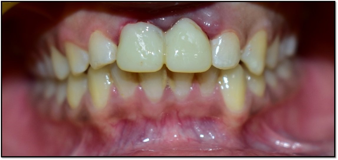
Figure 4: Temporization
Postoperatively, medicine was given 1 weeks. The patient was advised to adhere to a soft diet and avoid forces on the anterior teeth.
Patient was recalled after one and half month for prosthetic procedures and adequate amount of interdental papilla and the buccal contours were observed similar to the adjacent tooth, the abutment was removed and an impression coping placed, followed by a digital impression. The impression coping was removed and abutment was replaced. Digital impression was taken as an alternative to a conventional, rubber-based technique, a scan body was attached to the implant after removal of the provisional crown (Figure 5).
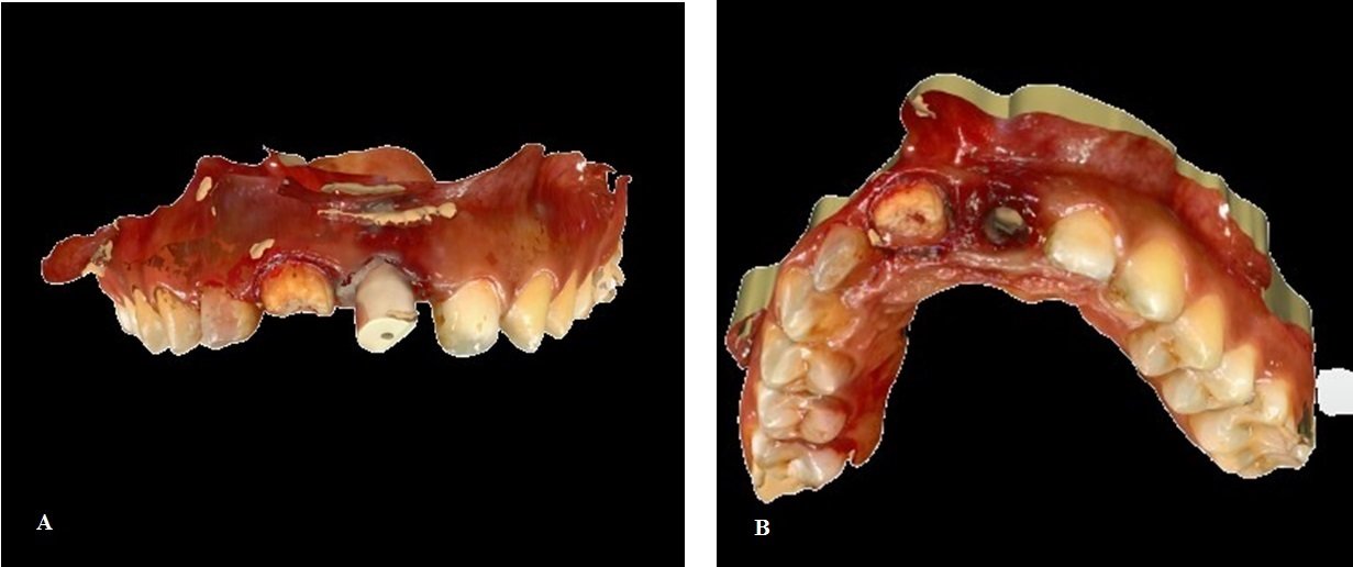
Figure 5 a,b: Digital impression
With a digital scanner capturing the position of the scanbody, a digital file was created, facilitating CAD/CAM fabrication of a custom zirconia abutment. The final crown, porcelain pressed to an all-zirconia core was seated on the final abutment. Then the final crown was cemented onto the custom abutment. Excess cement was removed and the occlusion was again verified. (Figure 6 a,b)
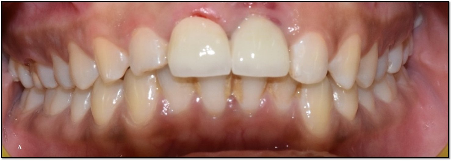

Figure 6 a, b: Occlusion was check
After 6 months, patient was recalled for follow up. The patient was very happy with the final aesthetic and functional outcome. Postoperative OPG was taken (Figure 7)
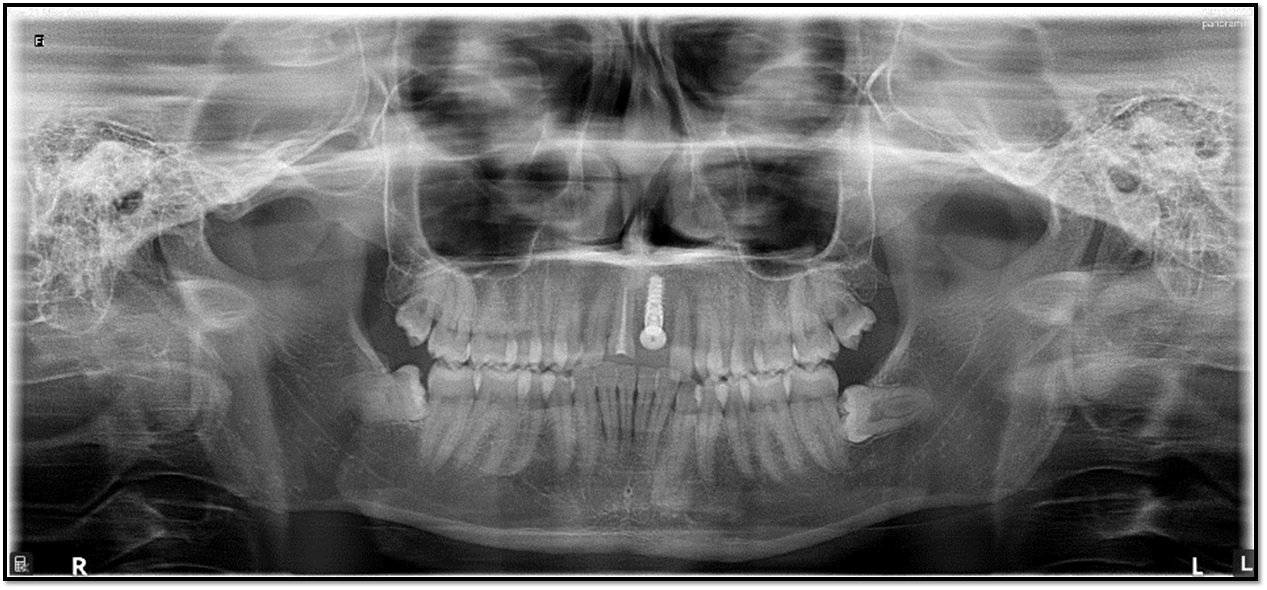
Figure 7: Follow up OPG
DISCUSSION
This case report discussed the key concepts of treatment planning, implant surgery, and prosthetic rehabilitation needed to achieve aesthetic success in the maxillary anterior region. The use of dental implants in the maxillary anterior region to replace missing teeth is a viable treatment option. There are many benefits of fixed dental implant-supported prosthetics versus traditional crown and bridge or removable tooth-borne prosthetics.16 Successful implant treatment to replace missing teeth in the anterior maxilla requires preoperative planning and a specific surgical plan, and ultimately prostheses are fabricated in consideration of function and soft-tissue support.17
Atraumatic extraction followed by implant placement in the anterior maxillary region is a challenge for dental clinicians. Precise implant placement in a 3D plane is the key for better aesthetics, function, and patient satisfaction. Immediate implant placement in upper anterior requires a thorough knowledge of anatomic, biologic, surgical, and prosthetic principles. Immediate placement of dental implants in the aesthetic zone is a technique sensitive procedure. However, immediate implant placement offers several advantages including preservation of alveolar bone, reduced treatment time, early prosthetic restoration with better aesthetics thus better patient acceptance. CBCT helps us to access all the required measurements in doing implantation. It provides highly accurate 3D images of the area concerned from a single, low-radiation scan, thus delivers a comprehensive detail of the patient’s jaw and the anatomical landmarks necessary to properly plan and provide treatment ensuring a better prognosis and outcome for implant placement. On the other hand occlusal adjustment, oral hygiene maintenance and regular recall check-ups should be considered prerequisites for maintaining a long lasting restoration.18
Decisive factors for the success of immediate implant are a lack of infection in periodontal tissues and an intact tooth socket. The evaluation of alveolar bone with CBCT helps the implantologist to assess the quality and quantity of cortical and cancellous bone, especially in the buccolingual dimension. CBCT has significantly contributed in the success of dental implant therapy in recent times. As a wide range of techniques in implant treatment is being applied, CBCT must be recommended as a diagnostic tool for implant treatment planning.19,20 This involves using CBCT in the diagnostic work up of a provisional dental implant treatment plan and then confirming and finally implementing it using appropriate software.21
CONCLUSION
Immediate implant placement and temporization, when properly indicated, has three main advantages: timing, biology, and prosthetics. Treatment time and number of surgical procedures are reduced compared to a delayed approach. From a prosthetic perspective, placing an implant-retained provisional crown on the day of surgery simplifies the temporization in the anterior area, allowing the patient to leave the practice on the same day with a fixed provisional.
REFERENCES
- Gotfredsen K. A 10-year prospective study of single tooth implants placed in the anterior maxilla. Clin Implant Dent Relat Res 2012;14(1):80-7.
- Adell R, Lekholm U, Rockler B, Brånemark PI. A 15-year study of osseointegrated implants in the treatment of the edentulous jaw. Int J Oral Surg 1981;10(6):387-416.
- Schwartz-Arad D, Chaushu G. The ways and wherefores of immediate placement of implants into fresh extraction sites: A literature review. J Periodontol 1997;68(10):915-23.
- Nir-Hadar O, Palmer M, Soskolne WA. Delayed immediate implants: Alveolar bone changes during the healing period. Clin Oral Implants Res 1998;9(1):26-33.
- Kan JY, Rungcharassaeng K. Site development for anterior single implant esthetics: The dentulous site. Compend Contin Educ Dent 2001;22(3):221-6, 228, 230-1.
- Brånemark P.I. Osseointegration and its experimental background. J. Prosthet. Dent. 1983;50: 399–410. doi: 10.1016/S0022-3913(83)80101-2. [PubMed] [CrossRef] [Google Scholar]
- Chrcanovic B.R., Albrektsson T., Wennerberg A. Dental implants inserted in fresh extraction sockets versus healed sites: A systematic review and meta-analysis. J. Dent. 2015; 43:16–41. doi: 10.1016/j.jdent.2014.11.007. [PubMed] [CrossRef] [Google Scholar]
- Chrcanovic B.R., Martins M.D., Wennerberg A. Immediate placement of implants into infected sites: A systematic review. Clin. Implant. Dent. Relat. Res. 2015;17((Suppl. 1)) :e1–e16. doi: 10.1111/cid.12098. [PubMed] [CrossRef] [Google Scholar]
- AlTarawneh S., AlHadidi A., Hamdan A.A., Shaqman M., Habib E. Assessment of Bone Dimensions in the Anterior Maxilla: A Cone Beam Computed Tomography Study. J. Prosthodont. Off. J. Am. Coll. Prosthodont. 2018; 27:321–328. doi: 10.1111/jopr.12675. [PubMed] [CrossRef] [Google Scholar]
- Lee S.R., Jang T.S., Seo C.S., Choi I.O., Lee W.P. Hard Tissue Volume Stability Effect beyond the Bony Envelope of a Three-Dimensional Preformed Titanium Mesh with Two Different Collagen Barrier Membranes on Peri-Implant Dehiscence Defects in the Anterior Maxilla: A Randomized Clinical Trial. Materials. 2021; 14:5618. doi: 10.3390/ma14195618. [PMC free article] [PubMed] [CrossRef] [Google Scholar]
- Schropp L., Wenzel A., Kostopoulos L., Karring T. Bone healing and soft tissue contour changes following single-tooth extraction: A clinical and radiographic 12-month prospective study. Int. J. Periodont. Restor. Dent. 2003;23: 313–323. [PubMed] [Google Scholar]
- Zhou Y., Si M., Liu Y., Wu M. Likelihood of needing facial bone augmentation in the anterior maxilla of Chinese Asians: A cone beam computed tomography virtual implant study. Clin. Implant Dent. 2019; 21:503–509. doi: 10.1111/cid.12787. [PubMed] [CrossRef] [Google Scholar]
- Fenner M., Vairaktaris E., Fischer K., Schlegel K.A., Neukam F.W., Nkenke E. Influence of residual alveolar bone height on osseointegration of implants in the maxilla: A pilot study. Clin. Oral Implant. Res. 2009; 20:555–559. doi: 10.1111/j.1600-0501.2008.01598. x. [PubMed] [CrossRef] [Google Scholar]
- Froum S.J. Immediate placement of implants into extraction sockets: Rationale, outcomes, technique. Alpha Omegan. 2005; 98:20–35. [PubMed] [Google Scholar]
- Kan J. Y., Rungcharassaeng K., Lozada J. Immediate placement and provisionalization of maxillary anterior single implants: 1-year prospective study. The International Journal of Oral & Maxillofacial Implants. 2003;18(1):31–39. [PubMed] [Google Scholar]
- Misch CE The importance of dental implants. Gen Dent. 2001;49:38-45.
- Tischler M. Dental implants in the esthetic zone. Considerations for form and function. N Y State Dent J. 2004;70:22-26.
- Novaes AB, Jr, Novaes AB. Immediate implants placed into infected sites: A clinical report. Int J Oral Maxillofac Implants. 1995;10(5):609–13. [PubMed] [Google Scholar]
- Van Assche N, van Steenberghe D, Guerrero ME, Hirsch E, Schutyser F, Quirynen M, et al. Accuracy of implant placement based on pre-surgical planning of three-dimensional cone-beam images: A pilot study. J Clin Periodontol. 2007;34(9):816–21. [PubMed] [Google Scholar]
- Mandelaris GA, Rosenfeld AL. The expanding influence of computed tomography and the application of computer-guided implantology. Pract Proced Aesthet Dent. 2008;20(5):297–305. [PubMed] [Google Scholar]
- Jung RE, Schneider D, Ganeles J, Wismeijer D, Zwahlen M, Hämmerle CH, et al. Computer technology applications in surgical implant dentistry: A systematic review. Int J Oral Maxillofac Implants. 2009;24(Suppl 92):109. [PubMed] [Google Scholar]
Disclaimer from Guident
The views and opinions expressed in articles published in Guident are solely those of the authors and do not necessarily reflect the views of the editors, the editorial board or the publisher, Ivory India. Guident does not endorse any product, technique or treatment described. Clinical content is intended for qualified dental and medical professionals and is not a substitute for individual clinical judgement. Guident, its editors and the publisher accept no liability for any consequence arising from the use of information published in the journal.
How to cite this article
Dr. Gaurav Gupta, Dr. D.K. Gupta, Dr. Devanshi Sankhla. Immediate Temporization in the Aesthetic Zone Using Noble Active Ti Ultra Implant: A Case Report. Guident. 2024 Feb;17(3). Available from: https://ivoryindia.in/articles/implantology/immediate-temporization-in-the-aesthetic-zone-using-noble-active-ti-ultra-implant-a-case-report.html