Implant Supported Prosthesis: A Case Report
Department-Conservative Dentistry and Endodontics, Department of Prosthodontics and Crown & Bridge, K.D. Dental College & Hospital, Mathura, UP, India.
INTRODUCTION
The goal of modern dentistry is to restore patients' oral health in a predictable manner. Depending on the patient's local surroundings, physiological state, emotional state, and financial situation, the best substitute tooth is used for missing teeth.1
It was defined in 1986 by the American Academy of Implant Dentistry as a contact formed between a normal remodeling bone and an implant without the interposition of nonbone tissue, entailing a sustained transfer and distribution of load from the implant to and within the bone tissue.2
A titanium screw with a smooth or roughened surface, like a tooth root, makes up a standard conventional implant. Commercially pure titanium, which comes in four grades based on the concentration of carbon, nitrogen, oxygen, and iron, is used to make the majority of dental implants.3
The main function of dental implants is to provide support for dental prostheses, or artificial teeth. The biological process by which bone bonds firmly to the surface of some materials, like titanium and some ceramics, is what gives rise to modern dental implants. Physical stresses can be supported by implants and bone integration for decades without experiencing any problems.4
It provides masticatory functions, and esthetics and restores the periodontal health of the patient. The treatment comprises of Surgical and Prosthetic Phase. The first phase is the Surgical phase comprising of placing the implant which is done by drilling the healthy bone and mimicking the root of the tooth. The Prosthetic phase is followed after 3-4 months of the treatment depending on the case.5
This case, report highlights the implant placement in the mandibular right second pre-molar region with screw-retained prosthesis.
CASE REPORT
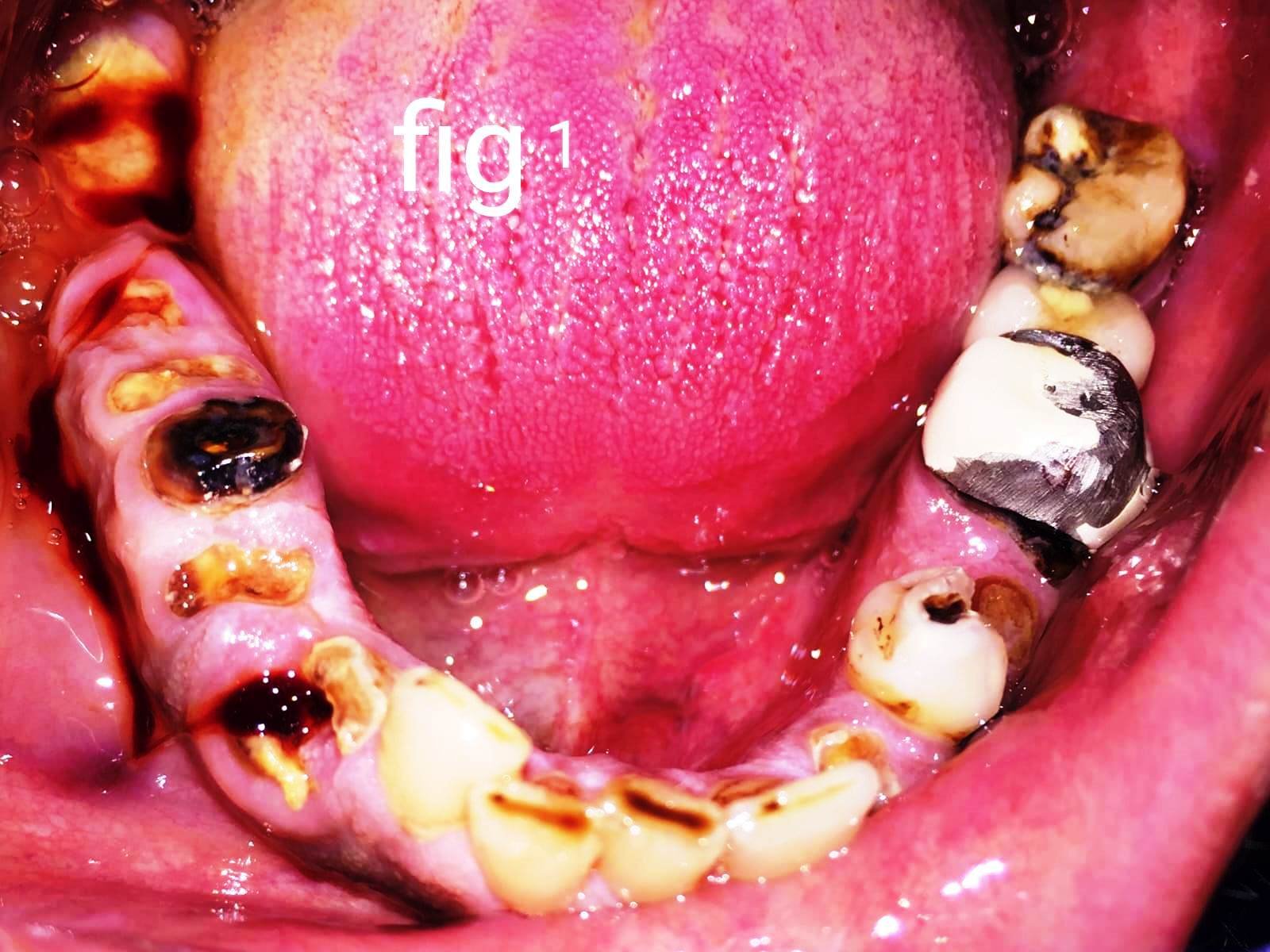
A 25-year-old Female patient reported to the Department of the Prosthodontic Crown and Bridge with the chief complaint of difficulty while chewing on the lower right back teeth region of the mouth due to missing teeth and unpleasant aesthetics due to a gap while smiling. She had undergone for extraction with the tooth four months ago as it was cariously destructed and hence wanted a replacement with the same. The patient was in good general health and she had no significant medical history. She has been explained all the treatment modalities for the replacement of the tooth that is removable and fixed prosthesis both tooth and implant supported along with a brief explanation of the pros and cons of the treatment. The patient decided to go for a fixed implant-supported prosthesis as she was well aware of the significant results of the same.
TREATMENT PLAN
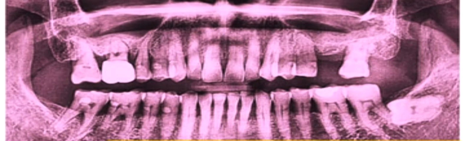
Preoperative OPG was advised which revealed significant bone height and width in 45 region. (Fig.1) Intraoral scanning was done to evaluate the occlusal height. CBCT was performed which gave an exact idea of the operatory site and its surrounding areas. The scan revealed that the bone type was D2. A super line dentium implant system was used. An implant size of 4.0x10.0mm was selected before the surgical procedure the patient was informed about the procedure and written consent was taken.
SURGICAL PROCEDURE
Once the patient was seated on the operating chair, first extraoral then intraoral scrubbing with povidone iodine, and then intraoral with 0.2% chlorhexidine digluconate mouthwash was done. The surgical site was anesthetized by local administration of 2% lignocaine hydrochloride with 1:80000 adrenaline by giving local infiltration of 1.5ml in the buccal and 1.5 lingual vestibule. The anesthesia was confirmed by subjective and objective signs of the patient. A midcrestal incision was made at the edentulous space (Fig 2).

Figure 1: Preoperative OPG

Figure 2: Midcrestal Incision
The mucoperiosteal flap was reflected (fig 3). The first drill was made with Lindman's guide then Lindman's first from dentium implant kit. (fig 4). An RVG was taken to evaluate the position of the drill. (fig 5)

Figure 3: Flap elevation

Figure 4: Dentium Implant kit

Figure 5: Drill position
Sequential drilling was done till 4.0x10.0mm and osteotomy was completed. (fig 6) The implant was placed at 30rpm speed and an adequate torque of 35 Ncm. (fig 7) RVG was taken immediately after the placement (fig 8) and the cover screw was placed. Interrupted sutures were given for flap closure with the help of silk 3.0 sutures. (fig 9)

Figure 6: Osteotomy preparation

Figure 7: Implant placement

Figure 8: Radiographic image of Implant placement

Figure 9: Suture placement
POST-OPERATIVE CARE
Post-operative medications were given. To promote wound healing the patient was advised to warm saline rinses for an initial 15 days. Post-operative instructions were to avoid any undue stresses on the surgical site. After 15 days sutures were removed.
SECOND-STAGE SURGERY
Three months following the patient's recovery, they were called back. To evaluate the osseointegration, RVG was acquired. After giving local anesthesia the cover screw was extracted along with the soft tissue covering it using a round bur (Fig. 10,11). A healing abutment was placed.

Figure 10: removal of soft tissue

Figure 11: removal of cover screw
After 15 min the tray was selected for open tray impression. Healing abutment was removed and an open tray impression coping was placed and tightened with a hex driver (Fig 13) Impression was taken by using light body and PVS putty impression material and loosening the coping.

Figure 12: placement of healing abutment

Figure 13: Placement of impression coping
After removal of the impression place the healing abutment over the implant in the patient's mouth. The implant analog was attached to the coping (Fig 14) and made the gingival mask and the impression was poured. After the removal of the cast place the coping and make a jig over the cast (Fig 15) and the trial was done in the patient's mouth. (Fig 16) The patient was recalled after 15 days.

Figure 14: Placement of lab analog

Figure 15: Jig over the cast

Figure 16: Jig trial
PROSTHETIC PROCEDURE
After 15 days a screw-retained metal ceramic crown was fabricated according to the selected shade of the adjacent teeth. The final restoration was checked in place with RVG which showed perfect fit. The patient was satisfied with the results. The prosthesis was screw retained the assembly was screwed intraorally. (fig 17,18)

Figure 17: screw-retained prosthesis

Figure 18: Post-operative picture
DISCUSSION
For a wide range of clinical applications, endosseous dental implant therapy is quickly taking the place of prosthetic standards of care; yet, despite its high success rate, endosseous implant therapy has not gained widespread public approval or use.6
Dental implants have become the most reliable and acceptable alternative for missing teeth. They are the nearest equivalent to natural teeth and therefore a useful addition in the management of patients who have missing teeth because of disease, trauma, or developmental anomalies. Alternate treatment options considered for this case were a removable partial denture which can contribute to alveolar bone loss, also it has a very low satisfaction rate. Another option is fixed partial dentures would have compromised adjacent teeth to a certain limit.5
Although reports of endosseous implant therapy in the mandibular parasymphyseal mandible have consistently shown a success rate of 95% or higher, endosseous implant therapy has not yet been used widely enough.6
Dental implant prostheses have been widely used to effectively replace missing teeth with a survival rate ranging from 93 to 95%.7
Multiple factors can dictate dental implant success or failure, including systemic health, history of periodontal disease, and quality, and quantity of hard and soft tissues.8 Dental implants positively affect the overall well-being of the patient including psychology.9
CONCLUSION
Achieving initial stability and maintaining functional loads is the aim of implant rehabilitation. The justification for implants longer than 10 mm was accepted for a long time since it increases the area available for osseointegration.
REFERENCES
- Pandey C, Rokaya D, Bhattarai B.P. Contemporary Concepts in Osseointegration of Dental Implants: A Review. BioMed Rese Inter (2022);1-11
- American Academy of Implant Dentistry. Glossary of implant terms. The J Oral Impl. (1986);12(2):284–294
- Arturo N. Natali. Dent Biom, (2003). ISBN 978-0-415-30666-9, pp. 69-87.
- Misch CE.Cont Impl Dent. (2007). St. Louis, Missouri: Mosby Elsevier.
- Kadam S, Tembhurne J, Gangurde A, Jaiswal N, Mittal S. Dental Implant Prosthesis, A Savior For Natural Teeth: A Case Report, J Dent and Medi Sci. (2023); e-ISSN: 2279-0853, (Vol 22); Issue 6 Ser.2 PP 19-23
- Wagenberg BD, Ginsburg TR (2001) Immediate implant placement on removal of the natural tooth: retrospective analysis of 1,081 implants. Comp Cont Educ Dent 22:399–404
- Pjetursson B. E., Heimisdottir K. Dental implants - are they better than natural teeth? Euro J Oral Sci. (2018);126(Suppli1):81–8 doi: 10.1111/eos.12543.
- Berglundh T., Armitage G., Araujo M. G., et al. Peri-implant diseases and conditions: consensus report of workgroup 4 of the world workshop on the classification of periodontal and peri-implant diseases and conditions. (2018) J Perio.89: S313–S31
- Dr. Sonawane P, Dr. Tembhurne J, Dr. Gangurde A. Fixed Implant Prosthesis: A Case Report. J Dent and Medi Scien (2023), e-ISSN: 2279-0853, p-ISSN: 2279-0861.Vol 22, Issue 7 Ser.8 PP 38-42
Disclaimer from Guident
The views and opinions expressed in articles published in Guident are solely those of the authors and do not necessarily reflect the views of the editors, the editorial board or the publisher, Ivory India. Guident does not endorse any product, technique or treatment described. Clinical content is intended for qualified dental and medical professionals and is not a substitute for individual clinical judgement. Guident, its editors and the publisher accept no liability for any consequence arising from the use of information published in the journal.
How to cite this article
Dr. Juhi Dubey, Dr. Manesh Lahori, Dr. Abhishek Sharma. Implant Supported Prosthesis: A Case Report. Guident. 2024 Feb;17(3). Available from: https://ivoryindia.in/articles/implantology/implant-supported-prosthesis-a-case-report.html