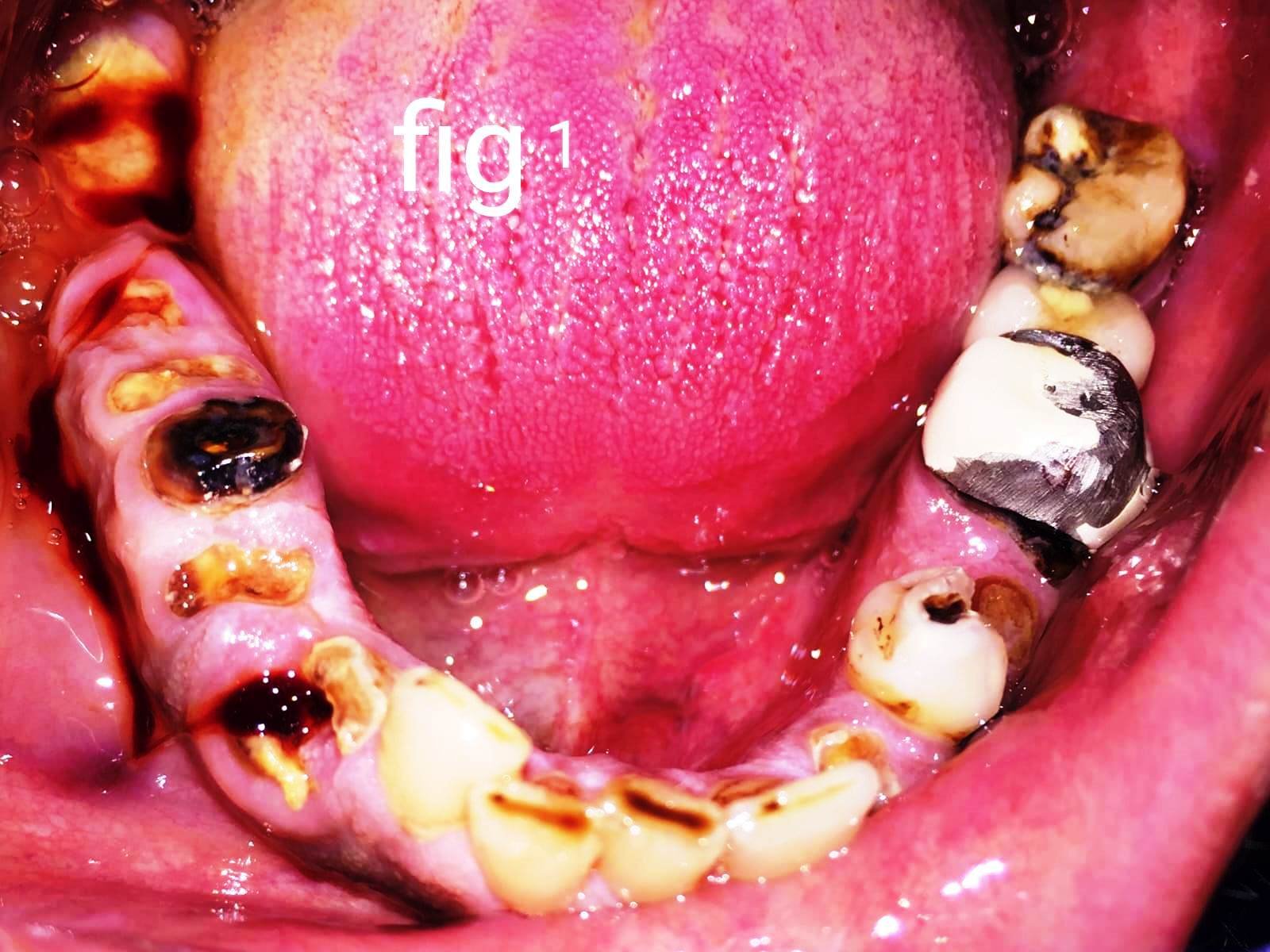
Tuberopterygoid Implants: Atropic Posterior Maxillary Rehabilitation Using Cortico Basal Implants: A Case Report
V. K. Dental, Bangalore, KAR India
ABSTRACT
The anatomy of maxilla is in such a way that rehabilitation with implants at posterior maxilla without putting up the cantilever in the prosthesis is sometimes very difficult because of various reasons such as pneumatization of maxillary sinus, failing grafting and sinus lift procedures, poor quality and quantity of bone. Here the alternative easier and accessible bone for the support with distal part of maxilla comes from the pterygoid fusion zone. Interestingly all the procedures are flapless, keyhole drilling, and immediate loading.
Possible because of specially designed implants with the cortical basal implants.
Key words; Strategic implantology, cortico basal implantology, basal bone, Osseo fixation, immediate functional loading strategy. pterygoid implant ,Tubero pterygoid implant
INTRODUCTION
While rehabilitating posterior maxilla meticulous planning and execution plays an important role. The axillary forces on pterygoid implant are well tolerated when they are joined with anterior most implants and reducing the span between anterior most posterior most pterygoid implant should be very less when rehabilitating the segments especially. The principle followed here is Osseo fixation with high primary stability and rigid stabilization hence the immediate loading.
CASE REPORT
A young man of age 65 having bruxism, close relative of my wife and diabetic had few teeth mobile with upper left quadrant An OPG revealed a bone loss with respect to upper left second molar and clinically grade 2 mobile and also bone loss with respect to upper left second premolar and a missing upper left first molar we planned to extract second premolar and second molar and planned immediate implant placement followed by immediately loading with long term Provisionals with 3d printing
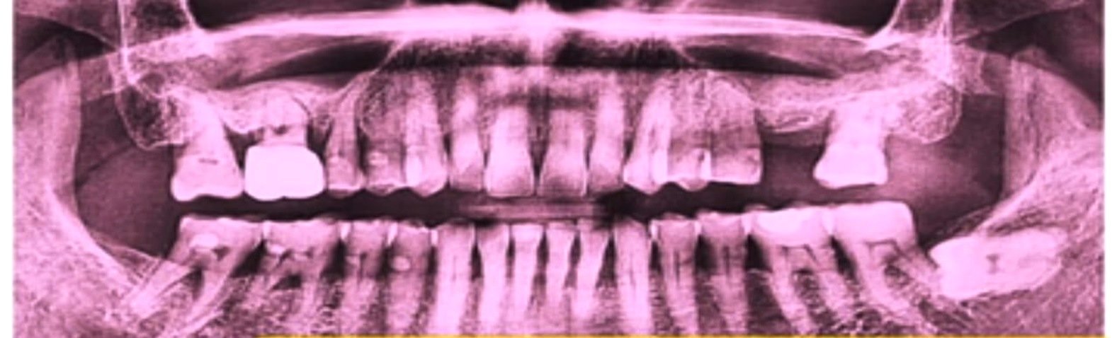
Fig 1

Fig 2
SURGICAL PHASE
After local anesthesia with lidocaine and posterior superior alveolar nerve block and infra orbital nerve block extraction of second premolar and second molar done povidine iodine irrigation and curettage and disinfection of socket done and on same appointment drilling made for implant placement without rising any flap and keyhole drilling.
First implant posterior to first premolar was engaged in the canine pillar area bypassing the root apex of first premolar and canine and the size of the implant used here is of company monoimplant 3.5mm diameter and length of 29 mm and posterior to that a wide body implant of 5.5mm of diameter with 8 mm of length is used to utilize the buccal palatal and sinus floor cortical and achieved more than 60 nm of torque and two pterygoid implant placed with the diameter and length of 3.5/ 23mm and 3.5/17mm all the implants were in high torque with at least of 60 nm.
All the implants were rigidly stabilized with 2 titanium wires of diameter 1.5mm with syncrystalization (intra oral welding).

Fig 3
PROSTHETIC PHASE
A direct digital scan made by intraoral scanner and using exocad the wax up has been made and printed with resin 3d printer with the help of resin called crown and bridge permanent from prevest denpro and insertion has been done dual cure luting resin cement on the same day.

Fig 4

Fig 5
ANATOMICAL CONSIDERATIONS AND RADIOGRAPHIC EVALUATION
A pterygoid implant placement is considered to be more successful treatment protocol followed an atrophy in the maxilla is concentric in nature the anatomical variations become complex to place a pterygoid implant anatomically being medial is safer than going laterally.
The vital structures u may encounter are the maxillary artery pterygo palatine fossa and injuring the pterygoid venous plexus happens when we enter lateral to the residual alveolar ridge.
The anatomical landmark which is felt intraorally is the medial pterygoid hamulus 3 to 4 mm lateral and slightly superior to it is our target area between the two pterygoid plates.

Fig 6

Fig 7
An implant other than the true pterygoid implant
- Implant in the pyramidal process of palatine bone,
- An implant in the posterior wall of maxillary sinus,
- An implant in the palatine bone considered to be successful treatment protocol with the condition of high primary stability of above 60 n m of torque
PARP (pterygoid anatomic radiographic prediction) Luis et al. proposed the classification of diagnostic prediction PARP for implantology in the pterygomaxillary region. Through the PARP, the choice of implant is individualized for each patient, acting as a guide to make implantology accessible in the pterygomaxillary region to the greatest number of specialists. From the degree of sinus invasion obtained after a three-dimensional (3D) computerized tomography (CT), the PARP establishes the prediction of the difficulty implied by implantology in this anatomical region, as well as the appropriate choice of the type of implant and length with which to approach it. The PARP classification allows working only in the pterygomaxillary region.
- PARP 1. It is the simplest scenario when there is no sinus invasion, and we have a bone in all its route. In these cases, the length of the implant depends on the bone density.
- PARP 2. The patient presents with a sinus invasion but still has >10 mm of the remaining bone. In case of having good bone density,
- PARP 3. This is a case of medium-high difficulty, with sinus invasion leaving a bone surface between 5 mm and 9 mm of remaining bone. In these cases, due to the scarce remnant of alveolar bone and the air of the sinus invasion, the pterygoid anchor will always be used in the apophasis of the same name, with a suitable density.
- PARP 4. In the majority of cases of a large sinus invasion, leaving only a remaining bone smaller than 5 mm, the possibility of using long pterygoid implants or opting for other surgical approaches will be evaluated

Fig 8
CONCLUSION
Extremely atrophic maxillae are the most challenging task for restorative dentists. Pterygoid implant provides a reasonable alternative to 3D maxillary reconstruction, sinus lifts, and bone augmentation technique. Many authors have reported success rates of pterygoid implants ranging from 90% to 100% after follow-up period ranging from 1 to 12 years with minimal complications.
Avoidance of a prosthetic distal cantilever with good stability fit for immediate loading is possible with this technique.
REFERENCES
- Prosthetic principles for corticobasal implantology by Prof, Dr Veerendrakumar . S .C
- Scortecci G. Basal Implantology. New York: Springer International Publishing, 2019. DOI: 10.1007/978-3-319-44873-2
- Ihde S, Ihde A, editors. Munich, Germany: The International Implant Foundation Publishing; 2011. Immediate Loading.
Disclaimer from Guident
The views and opinions expressed in articles published in Guident are solely those of the authors and do not necessarily reflect the views of the editors, the editorial board or the publisher, Ivory India. Guident does not endorse any product, technique or treatment described. Clinical content is intended for qualified dental and medical professionals and is not a substitute for individual clinical judgement. Guident, its editors and the publisher accept no liability for any consequence arising from the use of information published in the journal.
How to cite this article
Dr. Veerendrakumar SC., Dr. Shivakumar Kotturu. Tuberopterygoid Implants: Atropic Posterior Maxillary Rehabilitation Using Cortico Basal Implants: A Case Report. Guident. 2024 Feb;17(3). Available from: https://ivoryindia.in/articles/implantology/tuberopterygoid-implants-atropic-posterior-maxillary-rehabilitation-using-cortico-basal-implants-a-case-report.html