Hemifacial Microsomia: Face with Deficiency on One Side
Amrita School of Dentistry, Kochi, Kerala, School of Dental Sciences, Krishna Institute of Medical sciences, Karad, Maharastra., Mahe Institute of Dental Sciences, Mahe, Pondicherry, India.(Note: All 3 Authors had done these cases as PG Student in Yenopaya Dental College Mangalore), Department of Oral Medicine and Radiology
Abstract
Hemifacial microsomia is the second most common congenital facial anomaly of first and second branchial arches involving underdevelopment of one side of the temporomandibular joint, mandibular ramus, masticatory muscles, ears and occasionally defects in facial nerve and muscles. The most obvious deformity involves the lower jaw and ear, but soft tissue deficiency, maxillary hypoplasia and even orbit and skull anomalies may also be present. The deformities in hemifacial microsomia may vary greatly in extent and degree covering a wide spectrum ranging from mild underdevelopment of the lower jaw to severe deformity of the skull and face. Here, we report two cases of hemifacial microsomia diagnosed based on clinical and radiographic findings.
Keywords: Hemi facial microsomia, orthopantomogram, congenital facial anomaly.
INTRODUCTION
Hemifacial microsomia (HFM) is described as a spectrum of complex anomalies characterized by underdevelopment of one side of the face that results from a developmental malformation of the first and second branchial arches. HFM was first described by German physician Carl Ferdinand Von Arlt in1881.1 Hemifacial microsomia is also described by the names such as craniofacial microsomia, lateral facial dysplasia, first and second branchial arch syndrome and oculoauriculovertebral dysplasia.2 The occurrence of HFM is between 1 in 3000 and 1 in 5600 births.3 Males appear to be more frequently affected than females.4
Here, we report two cases of hemifacial microsomia, one a 12 year old female patient and another a 30 year old male patient which were diagnosed based on the clinical and radiographic findings.
CASE REPORTS
Case 1
A 12 year old female patient reported to our department with a chief complaint of asymmetry of right side of face. Her medical and family history were non-contributory. Her dental history revealed that she had undergone orthognathic surgery before 6years. Extraoral examination revealed underdevelopment of the right side of face (Fig 1). Her right eye was of smaller in size compared to contralateral side. The corner of the mouth on the left side was raised when compared to the other side. The ear on the same side was rudimentary without pinna, tragus and ear lobule (Fig 2). Surgical scar marks & screws were present at the region of right ear. TMJ examination revealed presence of deflection to right side (Fig 3). Her mouth opening was normal. Intra oral examination was not significant except for the presence of constricted maxillary arch with high arch palate seen. Orthopantomogram revealed a hypoplastic mandible and shortened ramus height and shortened size of both coronoid process and condylar head.(Fig. 4). The patient was referred to department of prosthodontics for an artificial ear prostheses and to the department of Oral surgery and plastic surgery for further treatments.
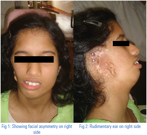

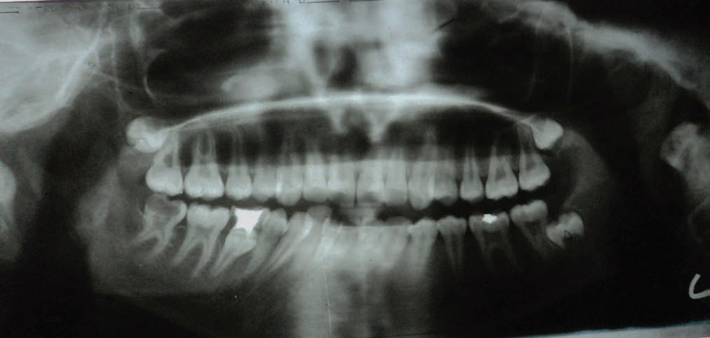
Fig 4- Orthopantomogram showing shortened condylar head & ramus height(on right side)


Fig 7- Orthopantomogram showing reduced ramus height & condylar head size (on right side)
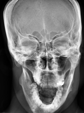
Fig 8- PA skull showing midfacial deficiency on right side
Case 2
A 30 year old male patient presented to our department complaining of underdevelopment of right side of face. Extraoral examination revealed underdevelopment of the right side of the face (Fig 5) with low set pinna and preauricular skin tags (Fig 6). Intra oral examination was not significant. The orthopantomogram showed an underdeveloped right side mandible with short ramus and shortened coronoid and condyle (Fig. 7). PA skull showed the presence of midfacial deficiency (Fig 8). The patient was referred to dept of Oral surgery and plastic surgery for treatments.
DISCUSSION
Hemifacial microsomia is the second most common congenital facial anomaly after cleft lip/palate in which there is deficiency in the amount of hard and soft tissues on one side of the face.5 It is primarily a syndrome of first and second branchial arches involving underdevelopment of the temporomandibular joint, mandibular ramus, masticatory muscles, ears and occasionally defects in facial nerve and muscles.1 In HFM, right side is affected more often than the left side with a ratio of 3: 2.6 Both the cases reported here are affected with HFM of right side.
Hemifacial microsomia is considered to be a developmental abnormality with an etiology of early loss of neural crest cells which is responsible for the clinical presentation of HFM.7 The clinical picture of HFM varies from slight asymmetry in the face to severe underdevelopment of one facial half with orbital implications. The most obvious deformity involves the lower jaw and ear, but soft tissue deficiency, maxillary hypoplasia and even orbit and skull anomalies may also be present. Lack of development of the external ear is a common feature with the severity of the ear deformity proportional to the jaw deformity. The chin and the facial midline are off-centered, and deviate to the affected side. Often, one corner of the mouth is situated higher than the other, giving rise to an oblique lip line.3 Hearing problems are present in 30-50% of patients.8< Intra-oral structures can also be affected in this condition. There will be agenesis of the third molar and second premolar on the affected side, as well as supernumerary teeth, enamel malformations, delay in tooth development and hypoplastic teeth. There will be hypoplasia of masseter, temporal and pterygoid muscles and the muscles of facial expression on the affected side. The degree of underdevelopment of the bone is directly related to the hypoplasia of the muscle to which they are attached.[3] In most cases, there is an underdeveloped condyle, but aplasia of the mandibular ramus and/or condyle with the absence of one glenoid fossa may also occur. In these cases, the maxilla is hypoplastic on the affected side as seen in our case also.
A panoramic radiograph provides an excellent overview of the osseous structures of the mandible and maxillofacial complex. A lateral cephalometric radiograph will help to evaluate the maxilla-mandibular relationship to the cranial base. PA view can be used to depict the degree of osseous asymmetry of the face. Computed tomography (CT) can provide both a three-dimensional rendition of the soft tissue of the face and an image of the underlying bone.7
The differential diagnosis of this condition includes Pierre Robin syndrome, Moebius syndrome and Treacher Collin’s syndrome. In cases of Pierre Robin syndrome, there will be cleft palate, micrognathia and glossoptosis. Moebius syndrome is a nonfamilial deficient development of cranial muscles consisting of facial diplegia with bilateral paralysis of the ocular muscles, particularly those supplied by abducens. HFM usually does not lead to ocular muscle paralysis and nerve involvement occurs unilaterally. Most of the features of Treacher Collin’s syndrome mimic HFM; however, the latter occurs unilaterally and it is sporadic in a vast majority of cases.9
In treatment planning, the dental occlusion must be considered with the underlying skeletal condition. In these patients, a combined surgical orthodontic approach is taken. Distraction osteogenesis is now widely accepted as a treatment modality for HFM. This is a gradual method of creating bone after a surgical corticotomy sectioning of the cortical plates. Prosthetic ear reconstruction can also be done for deformed ears as done in our case also.3
REFERENCES
- Dhillon M, Mohan SR, Suma NG, Raju MS, Tomar D. Hemifacial microsomia: a clinicoradiological report of three cases. Journal of Oral Science 2010; 52( 2): 319-324.
- Kalsotra P, Chowdhary A, Bhagat DR, Parihar SS, Prabhakar R .Craniofacial microsomia .J K Sci 2006; 8: 168-170.
- Moulin-Romse´e C, Verdonck A, Schoenaers J, Carels C. Treatment of hemifacial microsomia in a growing child: the importance of co-operation between the orthodontist and the maxillofacial surgeon. Journal of Orthodontics 2004;31:190–200.
- Cousley RR, Wilson DJ. Hemifacial microsomia: developmental consequence of perturbation of the auriculofacial cartilage model. Am J Med Genet 1992; 42: 461–6.
- Kapur R, Kapur R, Sheikh S, Jindal S, Kulkarni S. Hemifacial microsomia: a case report. J Indian Soc Pedod Prevent Dent 2008;26: S34-S40.
- Wang RR, Andres CJ. Hemifacial microsomia and treatment options for auricular replacement: a review of the literature. J Prosthet Dent 1999; 82: 197–204.
- Monahan R, Seder K, Patel P, Alder M, Grud S, O’Gara M. Hemifacial microsomia: etiology, diagnosis and treatment. J Am Dent Assoc2001; 132:1402-1408.
- Carvalho GJ, Song CS, Vargervik K, Lalwani AK. Auditory and facial nerve dysfunction in patients with hemifacial microsomia. Arch Otolaryngol Head Neck Surg 1999; 125: 209-212.
- Rajendran R, Sivapathasundaram B. Shafer’s textbook of oral pathology 2006; 5th ed, Elsevier, New Delhi: 986-989,1189-1190.
Disclaimer from Guident
The views and opinions expressed in articles published in Guident are solely those of the authors and do not necessarily reflect the views of the editors, the editorial board or the publisher, Ivory India. Guident does not endorse any product, technique or treatment described. Clinical content is intended for qualified dental and medical professionals and is not a substitute for individual clinical judgement. Guident, its editors and the publisher accept no liability for any consequence arising from the use of information published in the journal.
How to cite this article
Dr. Sreeja P Kumara, Dr. Suresh KV, Dr. Muhammad Shanavas. Hemifacial Microsomia: Face with Deficiency on One Side. Guident. 2014 Jul;7(8). Available from: https://ivoryindia.in/articles/oral-medicine/hemifacial-microsomia-face-with-deficiency-on-one-side.html