
Pocket of Teeth Brief Review of Literature, Differential Diagnosis and Management Options
(Prof. & HOD), Department of Oral and Maxillofacial Surgery, Nair Hospital Dental College, Opposite Maratha Mandir Theatre, Mumbai
ABSTRACT
Odontomas account for the most common odontogenic tumours which are often asymptomatic and discovered on routine radiography. They are also known to cause eruption disturbances necessitating its surgical excision as the viable option. They are considered as “hamartomas” rather than true neoplasms and consist of tooth components bearing little or no resemblance to the teeth. We present this case series of 8 such cases to report our wide experience in managing this condition with a review of literature and differential diagnosis.
Keywords: Odontoma, Compound, Complex, Mandibular
INTRODUCTION
The term ‘odontoma’ or ‘odontome’ was firstly described by Paul Broca in 1867 which was then used as a general descriptive term for any tumour (in its broadest sense) of odontogenic origin. Odontoma is considered to be hamartomatous malformations rather than true neoplasms and is the most common odontogenic tumour comprising of 22% of all odontogenic tumours1. They are composed of the tissues native to teeth: enamel, dentin, cementum and pulp tissue1. They are considered as a growth in which both the epithelial and mesenchymal cells undergo complete differentiation, with the result that functional ameloblasts and odontoblasts form enamel and dentin, respectively. This enamel and dentin are usually laid down in an abnormal pattern as the organisation of the odontogenic cells fails to reach a normal state of morphodifferentiation2. As this lesion consists of more than one type of tissue, it is also termed as ‘composite odontoma’.
According to the WHO Classification (2005), they have also been termed as ‘Compound’ and ‘Complex’ Odontomes based on the degree of anatomic similarity to the native teeth. The term ‘compound odontome’ is used when there is at least some degree of resemblance to the normal teeth. ‘Complex’ Odontome’ is composed of irregular calcified mass bearing no morphological similarity even to rudimentary teeth.2.
The odontomas are generally small, slow growing and asymptomatic lesions which with continued growth show late bony expansion. The lesions seldom cause any clinical signs or symptoms and are frequently discovered on routine radiographic examination. However few cases3 of odontomas causing pain have been reported. At times the lesions also are seen to cause some pathological changes such as malposition, delayed eruption, impaction, cyst formation and displacement/resorption of the adjacent teeth involved.
They are seen to occur predominantly in the first two decades of life with a slight female predilection.4
The highest majority of odontomas occurring in the anterior maxilla are the ‘compound composite’ types, whereas those occurring in the posterior region, especially, of the mandible are the ‘complex composite’ type2. Rare occurrence of such lesions has been reported in the maxillary sinus and subcondylar regions5.
The etiology of odontomas still remains unclear. There are many speculations made regarding the etiology. These lesions are the rarest to show recurrence and are treated by simple conservative surgical excision. In case of recurrence, ruling out other odontogenic lesions like calcifying odontogenic cyst and ameloblastic fibrodontoma are crucial6. We present a case series of compound and complex odontomas occurring in adult and paediatric population with a brief review of literature and differential diagnosis.
CASE REPORTS
Case 1
A 13 year old child reported to the OPD with the complaint of pain and mild swelling in the right anterior region of mandible since 6 months. There was no previous history of similar symptoms reported. The medical history was non-contributory. General examination did not reveal any abnormality.
Extraoral examination did not reveal any local swelling/ tenderness/ abnormality (fig 1). Intraoral examination showed slight labial cortical expansion in the right anterior mandible. Remarkably, the patient was also seen to have over retained deciduous teeth 81, 82 and 83. The swelling was well-defined and roundish which elicited mild tenderness on palpation. There were no other significant local changes (fig 2).

Fig 1: Pre-op extraoral profile view

Fig 2: Pre-op intraoral view showing labial cortical expansion in right anterior mandible and over-retained deciduous 81, 82 and 83.
Panoramic radiograph revealed calcified mass in the anterior mandible with impacted 42 and 43 (fig 3). CBCT examination revealed a conglomerate mass of tooth like structures in the right mandibular anterior region causing thinning and expansion of labial cortical plate and impeding eruption of the adjacent teeth (fig 4a to c).

Fig 3: Panoramic radiograph showing conglomerate mass of calcified structures in the anterior mandible with impacted 42 and 43

Fig 4a

Fig 4b
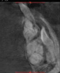
Fig 4c
Fig 4a to c – CBCT scans showing a mass of calcified tooth like structures causing thinning and perforation of labial cortical plate.
The patient was diagnosed with ‘Compound Composite Odontoma’ based on clinical and radiographic examination and was accordingly taken up for the excision procedure under LA. Crevicular incision was made from canine to canine with releasing incisions on either side and a trapezoidal mucoperiosteal flap was reflected. Labial cortical thinning and expansion was evident. Further de roofing of the labial cortex was done and pathology was exposed. The entire calcified mass was removed in toto with its capsule, while taking care not to damage the associated impacted teeth. Hemostasis was achieved. The resultant bony cavity was smoothened. The site was flushed with Normal saline + Povidone Iodine solution. Closure was performed using 3-0 VICRYL sutures (fig 5a to c). The mass was further submitted for histological examination.

Fig 5a

Fig 5b

Fig 5c
Fig 5 a to c – Deroofing of the calcified mass followed by the resultant surgical defect and excised mass.
Postoperative panoramic radiograph was made which showed complete excision of the calcified mass (fig 6). Histopathology confirmed the provisional diagnosis of ‘Compound Composite Odontoma’. Postoperative healing was uneventful and the patient was free from his symptoms. The patient was explained about the need for orthodontic treatment and/or second surgery to facilitate eruption of the impacted teeth and was also kept on a regular follow up.

Fig 6: Post-operative panoramic radiograph

Fig 7a

Fig 7b
Fig 7a to b: Post operative extraoral and intraoral images. Satisfactory intraoral healing is evident.
Case 2
A 15 year old male reported to the Maxillofacial surgery OPD in view of missing teeth in the upper anterior region of the jaw (fig 8). No previous history of trauma or dental extraction was reported. The patient was examined and was found to have a missing left central incisor along with a normal compliment of teeth (fig 9).

Fig 8: Pre-op extraoral profile view

Fig 9: Pre-op intraoral view showing missing 21 and no obvious bony expansion

Fig 10- Panoramic view showing a conglomerate mass with impacted supernumerary and 21
On panoramic radiographic a calcified mass was seen in the anterior maxilla with an impacted supernumerary tooth lying above it and impacted 21 close to the nasal floor (fig 10).
Additionally, the CBCT revealed thinning of the left lateral nasal wall caused by the calcified mass and presence of labially tipped impacted supernumerary plus horizontally impacted 21 above the calcified mass and the supernumerary tooth close to the nasal floor (fig 11a and 11b). The calcified mass was seen to have a morphological resemblance to normal teeth and was thereafter diagnosed as a ‘Compound Composite Odontoma’.

Fig 11a

Fig 11b
Fig 11a to 11b: CBCT sections showing a well-defined calcified mass with resultant thinning of the lateral nasal wall and the presence of impacted supernumerary and horizontally placed 21.
The patient was referred to Dept. of Orthodontics for opinion regarding the prognosis of impacted 21, and was declared to have a poor orthodontic prognosis. Hence a surgical removal of the calcified mass, the supernumerary tooth and the impacted 21 was planned under GA.
Accordingly, the patient was taken up for the procedure, a trapezoidal mucoperiosteal flap was reflected. Labial cortical thinning and expansion was evident (fig 12). Further de roofing of the labial cortex was done and the pathology was exposed (fig 13). The entire calcified mass was removed in toto with its capsule. The supernumerary and the impacted 21 were subsequently retrieved (fig 14). Hemostasis was achieved. The resultant bony cavity was smoothened. The site was flushed with Normal saline + Povidone Iodine solution. Closure was performed using 3-0 VICRYL sutures (fig 5a to c). The mass was further submitted for histological examination.
Histopathology confirmed the provisional diagnosis of ‘Compound Composite Odontoma’. Postoperative healing was uneventful and the patient was free from his symptoms.


Fig 12: Intraop view of the expanded labial cortex

Fig 13: Deroofing of the cortex and exposure of the calcified mass

Fig 14: Excised mass with its fibrous capsule, extraction of impacted supernumerary and 21.
DISCUSSION
Odontomas are considered as benign hamartomatous malformations having a slow course of growth while causing no frank symptoms. They are the most common odontogenic tumours and constitute 22% of all the odontogenic tumours of the jaws.2
Odontomas are commonly seen in the first two decades of life with a predilection for occurrence in the female gender. The incidence of Compound Odontomas ranges somewhere between 9% to 37% where majority of them occur in the anterior maxilla (61%), whereas a majority of the odontomas found in the posterior jaw regions, especially the mandible are of the ‘Complex’ type.2, 6, 7 Interestingly, in our series of patients we noted similar findings in terms of age, along with a high prevalence of ‘Compound Odontomas’ in the anterior maxillae and ‘Complex’ ones in the posterior jaw regions.
Reports published also have shown that odontomas more commonly occur in the right side of the jaws as compared to the left2, 7 which is interestingly true for our series of cases.
Moreover ‘Compound’ variety occurs commoly in the incisor-cuspid area2, 7 and the ‘Complex’ type in premolar-molar regions of the jaw2, 7 which has a striking resemblance to our case series too.
The etiology still remains unclear. It is thought to develop following a local infection/ trauma. Also it has been seen to be associated with inflammatory and infectious processes, hereditary anomalies (Gardner’s syndrome, Hermann’s syndrome), hyperactivity of odontoblasts and alterations in the genetic components responsible for control of tooth development postnatally2, 6.
Clinically, the odontomas are intraosseous (central), peripheral (extraosseous or soft-tiisue) and erupted odontomas. The central variety is most common (51%) which occurs either as a ‘Compound’ (most commonly in the anterior maxilla) or ‘Complex’ (most commonly in the posterior regions) type.7, 8
The peripheral ones are quite rare and reported cases have presented in the soft tissues over the tooth bearing areas, and were surprisingly found to be of the ‘Compound’ type.8
The last variety of odontomas known as the ‘Erupted Odontoma’ usually presents coronal to the crown of an unerupted or an impacted tooth or lying superficially in the bone enabling its eruption.7 The mechanism of eruption of the ‘Erupted Odontoma’ is different from a normal tooth due to lack of periodontal ligament, and can be attributed to sequestration of the overlying bone/ remodelling of the jaws gradually causing occlusal movement.7 All our reported cases were of the ‘Central’ (intraosseous) variety; however we did not find any ‘Peripheral’ or ‘Erupted’ odontomas in our population.
Odontomas reported in the extragnathic tissues are pretty rare, with only 9 cases involving the nasopharynx, tympanic membrane, middle ear and brain reported so far in literature.9, 10 It has also been postulated that there are embryological alterations occurring within the primordial stomodeum and Rathke’s pouch11 which explains the occurrence of odontogenic tumour in a distant site.
The reliable way to clinically differentiate the complex and compound types of odontomas is through radiographic examination. The compound variety appears as a well-organized mass containing tooth like structures bearing resemblance to normal or rudimentary teeth; whereas the complex type appears as an irregularly arranged oval shaped radiopacity with no resemblance and usually surrounded by a narrow radiolucent rim2, 4 resembling the connective tissue capsule histologically.
Microscopic examination of both compound and complex odontomas for reaching to a definitive diagnosis cannot be overemphasised. The radiological appearances progress from radiolucent lesion initially followed by partial calcification in the intermediate stage and finally radiopaque amorphous appearance in the third stage.6
According to Kaban, the odontomas are usually separated by a capsule, hence are easily enucleated and seldom cause any trauma to the adjacent teeth that are displaced by the lesion.12
Owing to the limited growth potential, we seldom find odontomas causing large expansions and resultant dentoalveolar or facial asymmetry. However, they should be addressed as they contain various tooth formulations that may predispose to pathologic (cystic) changes6, 7 as described earlier.
Although these lesions are considered painless, literature still reports odontomas causing local pain, paresthesia of lower lip, headache in the frontal area3 as well. This is however in contrast to the findings in our series where 5 cases reported initially with pain.
In case of a mixed radiolucent- radiopaque appearance, differentiation from adenomatoid odontogenic tumor (AOT), calcifying epithelial odontogenic tumor (CEOT), ameloblastic fibrodontoma, odontoameloblastoma should be made. Histological evaluation for presence of disorderly arrangement of well- formed dental tissues can give us a clue.
These lesions never pose a challenge for diagnosis based on histology as the lesion is routinely composed of enamel, dentin and pulp tissue. The mature enamel is however expected to be lost during decalcification process and doesn’t appear on the routine H & E sections.
As this lesion is considered a non-recurrent one, it should be borne in mind that in case of an odontoma lying in close relation to an adjacent impacted tooth, removal of some part of the tumor can still be compromised which will prevent the risk of exposure and damage to the impacted tooth. Also, in case of a large mass, the tumor should be removed after sectioning to prevent use of undue forces for removal and damage to the surrounding structures like tooth/root/nerve(s)/nasal floor/nasal wall (s).
CONCLUSION
Recognition of the fact that complex odontomas can be seen in association with impacted or missing teeth is of utmost importance and can be favoured by timely evaluation of the panoramic radiograph and CBCT, thus preventing any further adverse effects. Thus, a thorough clinical and radiographic examination is mandatory to search for such lesions when clinical findings of a missing tooth/impacted or unerupted tooth are present. Early recognition and removal will help us to prevent complex treatments in future, risk of devitalisation and displacement of associated teeth and also to achieve a good prognosis.
CONFLICTS OF INTREST
None
REFERENCES
- Budnick SD (1976) Compound and complex odontomas. Oral Surg Oral Med Oral Pathol 42(4):501–506.
- Shafer WG, Hine MH, Levy BM. A textbook of oral pathology. 3rd ed.Philadelphia: WB Saunders Co; 1974: 276−8.
- Tu¨zu¨m MS (1990) Orofacial pain associated with an infected complex odontome case report. Aust Dent J. 35(4):352–354
- Nelson LB, Thompson DRL. Compound odontoma. Head Neck Pathol 2010; 4: 290–1.
- De Visscher JG, Gu¨ven O, Elias AG (1982) Complex odontoma in the maxillary sinus. Report of 2 cases. Int J Oral Surg 11(4): 276–280
- Reichart PA, Philipsen HP. Compound odontoma. In: Odontogenic tumors and allied lesions. London: Quintessence; 2004: 149−53.
- T.D.Daley, G. P.Wysocki, G.A. Pringle. “Relative incidence of odontogenic tumors and oral and jaw cysts in a Canadian population,” Oral Surgery, Oral Medicine, Oral Pathology, vol.77, no. 3, pp. 276–280, 1994.
- F. Ide, T. Shimoyama, and N. Horie, “Gingival peripheral odontoma in an adult: case report,” Journal of Periodontology, vol. 71, no. 5, pp. 830–832, 2000.
- McClatchey KD, Hakimi M, Batsakis JG: Retrotympanic odontoma. Am J Surg Pathol 5: 401e440, 1981
- McClure: Odontoma of the nasopharynx. Arch Otolaryngol 44: 51e60, 1946
- Bellucci RJ, Zizmor J, Goodwin RE: Odontoma of the middle ear. A case presentation. Arch Otolaryngol 101: 571e573, 1975
- L. B. Kaban and M. J. Troulis, “Dentoalveolar surgery,” in Pediatric Oral and Maxillofacial Surgery, p. 140, WB Saunders, Philadelphia, Pa, USA, 2004.
Disclaimer from Guident
The views and opinions expressed in articles published in Guident are solely those of the authors and do not necessarily reflect the views of the editors, the editorial board or the publisher, Ivory India. Guident does not endorse any product, technique or treatment described. Clinical content is intended for qualified dental and medical professionals and is not a substitute for individual clinical judgement. Guident, its editors and the publisher accept no liability for any consequence arising from the use of information published in the journal.
How to cite this article
Dr. Neelam N. Andrade Dean, Dr. Harsh K. Desai, Dr. Trupti Gandhewar, Dr. A. L. Nair Road. Pocket of Teeth Brief Review of Literature, Differential Diagnosis and Management Options. Guident. 2020 Jan;13(2). Available from: https://ivoryindia.in/articles/oral-surgery/pocket-of-teeth-brief-review-of-literature-differential-diagnosis-and-management-options.html


