Genetics and the Royal Deformity Class III: A Narrative Review
Department of Orthodontics, Karnavati School of Dentistry, Gandhinagar, Gujarat India, Department of Prosthodontics, College of Dental Sciences and Research Centre, Ahmedabad, Gujarat
INTRODUCTION
Who sees the human face correctly: the photographer, the mirror or the painter? ORTHODONTIST. Growth of the child especially the face is influenced by a lot many genetic and epigenetic factors which may turn out to be either favourable or unfavourable. Alexander Den Heijer rightly said, ‘When a flower doesn’t bloom, you fix the environment in which it grows, not the flower. In orthodontics, we follow the same principle. Orthodontics and Dentofacial Orthopaedics modify the environment of child’s facial growth by growth modification and after the growth, completion beautifies an individual either with camouflage or a surgical procedure by subsequently enhancing or maintain the function.
Malocclusion is a departure from normal occlusion. Edward H Angle classified malocclusion in 1899 based on antero-posterior jaw relationship as Class I, II & III malocclusion.1,2 Salzman explained the prenatal and postnatal causes of malocclusion. According to him the prenatal cause for malocclusion can either be genetic or congenital and post-natally it can either be genetic, functional and environmental.3 Moyers in 1972 mentioned seven etiologic factors causing malocclusion: Heredity, developmental defects, trauma, physical agents, habits, diseases and malnutrition.4Pitutary gland is the master gland which plays an important role in prognathism. Excessive growth hormone along with genetic influence makes prognathism more complex.
Class III Malocclusion is also referred as Mesio-occlusion, pre-normal occlusion, mandibular prognathism, Hapsburg jaw, and syndrome prognathiquemandibulaire (Jean Delaire). In OMIM (Online Mendelian Inheritance in Man) the term malocclusion reveals more than 100 entries for a variety of syndromes that includes malocclusion as one of their features. The best-known example is the familial “MP” (Mandibular Prognathism) (OMIM*176700) referred to as the Hapsburg jaw.5
Mandibular prognathism and Class III malocclusion are not synonyms (Guyer et al). Mandibular prognathism (Greek derivation where pro =forward and gnathos =jaw). There are two possibilities i.e the mandible is true prognathic in relation to the cranial base and maxilla or the mandible mimics prognathism because of either short cranial base or a deficient midface. Class III malocclusion is when the mesiobuccal cusp of upper permanent first molar occludes on the embrasure between the lower permanent first and second molar. Malocclusion is a dentoalveolar entity and it should never be used in synonyms with the jaw discrepancy. Thus, a dentoalveolar Class III malocclusion may or may not be superimposed on a skeletal Class III jaw base.
According to Angle (1899): Class III malocclusion occurred when the lower teeth occluded mesial to their normal relationship by the width of one premolar or even more in extreme cases. (Mesio-occlusion)(Figure-1). Vertical and transverse dimensions of the face were not considered. This classification addresses dental parameters and has no correlation with the maxillary and mandibular skeletal bases.2
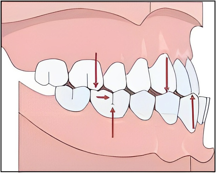
Figure 1: Class III Malocclusion
Class III malocclusion may often be present in mandibular prognathism, but it comprises only a part of a much larger syndrome. True mandibular prognathism is a skeletal imbalance which most often accompanies a Class III dental occlusion. The components of mandibular prognathism and Class III anteroposterior skeletal imbalances are:
- A large mandible relative to the maxilla
- A small maxilla relative to the mandible
- The maxilla is retropositioned to the mandible
- A forward rotation of mandible relative to cranium will cause the chin point to move into a more horizontally and protrusive position leading to a reduction in lower anterior facial height.6
Thus, the Class III malocclusion appear as a disharmonious relation between the maxilla and mandible. The morphological features of this group of malocclusions are variable and may include several craniofacial structures including an acute mandibular plane angle, an obtuse gonial angle, an overdeveloped mandible, an underdeveloped maxilla, a deficient orthocephalisation.
A wide range of environmental factors has been suggested as contributing to the development of Class III malocclusion. Among those are enlarged tonsil, difficulty in nasal breathing, congenital anatomic defects, disease of the pituitary gland, hormonal disturbances, a habit of protruding the mandible, posture, trauma and disease, premature loss of the sixth-year molar and irregular eruption of permanent incisors or loss of deciduous incisors.4
1.1 Sexual Dimorphism
Concentrations of sex hormones during pubertal growth spurt influences the craniofacial growth of both male and female differently. A high testosterone/oestrogen (T/E) ratio in puberty expedites facial features such as the lateral growth of mandible and chin, and the lengthening of the lower face.
Class III malocclusion shows a definite degree of sexual dimorphism after the age of 13. Females have a smaller linear dimension in maxilla, mandible and anterior facial height compared to males during circum-pubertal and post-pubertal periods. Males have relatively larger mandible and female have more hyperdivergent pattern of growth.7
1.2 Prevalence of Class III Malocclusion
Prevalence of class III malocclusion has a strong influence of ethnicity. The highest prevalence observed in East Asian populations such as Korean, Chinese, and Japanese (8%-40%).8 By comparison, African populations exhibit a reduced Class III prevalence rate (3%-8%) compared to Asians and Class III is relatively rare in individuals of European-American descent (0.48%-4%). Additional studies in the United States of America (US) have suggested that the prevalence of Class III malocclusion in Caucasians can be as high as (5.5%-9.5%)9, while the prevalence in US Native American Indian populations is relatively low (2.6%-3.1%).10
2 GENETICS AND CLASS III
Alison plowden says that the laws of genetics apply even if you refuse to learn them. Genetic influence on facial features is so strong that we can recognize many familial tendencies at a glance. Similarly, malocclusions are also inherited.
There are two major possibilities:
- Tooth size and Jaw size discrepancy
- Another likelihood is inheritance of disproportional size of upper and lower jaws, which would cause improper occlusal relationships.

Malocclusion of genetic origin includes the entire facial outline, prognathism, macro and micrognathia, abnormality in tooth size, arrangement and eruption, cleft lip and palate, facial clefts and bimaxillary protrusion and atresia.11
2.1 Mode of inheritance
The familial nature of mandibular prognathism was first reported by Strohmayer in 193712 and then Wolff et al in 199313 by pedigree analysis of Hapsburg family. Prognathic lower jaw, protruding lower lip as well as a dorsal hump prominence of nose (Hapsburg nose) were unique to the family. Multifactorial inheritance with autosomal recessive trait was noticed in this ‘Royal’ family because of extensive consanguinity.14, hence ‘The Royal Deformity’.
Suzuki studied offspring of parents with mandibular prognathism from 243 Japanese families, and reported a frequency of 31% of occurrence from paternal side, 18% from maternal side and 40% if both parents were affected.15 Nakasima et al assessed the role of heredity in the development of Angle’s Class II and Class III malocclusions and showed high correlation in the Class II and Class III groups. However, the role of cranial base, the midfacial complex and the mandible in the development of class III malocclusion has not been clarified yet.16 Saunders et al compared parents with offspring and siblings in 147 families and demonstrated a high level of significant correlations between first-degree relatives.17
Lobb suggested that the shape of the mandible and cranial base are more variable than the maxilla or cranium.18Manfredi et al found strong genetic control in vertical parameters and in mandibular structure in twins.19
Fernex et al found that the sizes of the skeletal facial structures were transmitted with more frequency from mothers to sons than from mothers to daughters.20 Hunter et al reported a strong genetic correlation between fathers and children, especially in mandibular dimensions.21
Most appropriate explanation for mode of inheritance was given by Litton et al in 1970 that Class III has polygenic mode of transmission which depends on the individual’s threshold to susceptibility. X chromosome does play a role in some syndromes with mandibular prognathism, the Class III phenotype is not X-linked since the numbers of affected males and females are similar.22
Genetic linkage studies have concluded that Class III tendencies have multiple genetic loci on different pair of chromosomes. Genetic epistasis is the feature of this malocclusion which means that the genetic configuration is influenced by many phenotypic variables.
2.2 Genetic component
Condylar cartilage is an important growth site in the mandible. The condylar cartilage is a secondary cartilage and supports the growth of the mandibular bone. Mechanical loading or functional stimuli might influence the responses of the condylar cartilage and the subsequent growth of the mandible.
McNamara and Carlson hypothesized that the cartilage of the mandibular condyle is responsive to biophysical environmental changes, and it is highly likely that Class III malocclusion might be precipitated under these biomechanical conditions by the inheritance of genes that predispose to a Class III.23 A number of reports have documented the influence of various genes that are involved in mandibular development. Growth factors and cytokines are local mediators and can be secreted in response to mechanical loading. These mediators regulate cell proliferation and the expression of differentiation products by activating signal transduction pathways in the target cells. In an experimental model of enhanced condylar growth, Rabie et al. indicated that forward positioning of the mandible triggered the expression of Indian hedgehog (Ihh) and Parathyroid hormone - like peptide (Pthlh), which promote mesenchymal cell differentiation and proliferation, respectively, and that these proteins acted as mediators to promote increased growth of the cartilage.24,25 Another rat model study by Hajjar D et.al found that the expression of IGF-1 increased significantly when the mandible was repositioned by means of a propulsive appliance. In addition, growth factors such as VEGF(Vascular endothelial growth factor) and transcription factors such as the sex determining region Y (SRY)-box 9 (Sox9) and Runt related transcription factor 2 (Runx2) play important roles in the differentiation of chondrocytes in the growth plate under conditions of mechanical loading or exposure to other stimuli.26 The genes that have been implicated in condylar growth by studies in the mouse might serve as potential candidates to increase our understanding of Class III malocclusion in humans.
Studies in mice also support the genetic basis of maxillary and mandibular size. Some investigators have used inbred mouse strains to confirm the hypothesis that the D12mit7 segment on mouse chromosome 12 determines maxillary growth27, while others applied Quantitative Trait Locus (QTL) studies in inbred mice to identify specific QTLs on mouse chromosomes 10 and 11 that are correlated with the anteroposterior length of the mandible.28
The Hox families of genes are master regulatory genes shown to play a definitive role in patterning the hindbrain and branchial regions of the developing head, including structures derived from the second branchial arch. The Hox3 region contains at least 7 genes in a 160-Kb stretch of DNA, including
Hoxc4, Hoxc5, Hoxc6, Hoxc8, Hoxc9, Hoxc10, Hoxc11, Hoxc12, and Hoxc13.29
The COL2A1 (collagen, type II, alpha 1) gene, located between positions 12q13.11 and12q13.2, encodes the alpha-1 chain of type II collagen found in cartilage. Previous studies in mice point confirms the importance of Col2A1 in craniofacial growth. Results from mouse studies of craniofacial growth show that a region on chromosome 12 is biologically relevant to craniofacial development and may be linked to the Class III phenotype.30
In an SMXA recombinant inbred strain of mice, the positions of the mouse chromosome 10 and chromosome 11 were determined to be responsible for mandibular length and corresponded to regions 12q21 and 2p13, respectively, in human chromosomes. These results suggest that the major genes responsible for mandibular length are in these regions. Progress in the craniofacial genetics field toward human genetic mapping of the Class III trait is gradual but limited.28
3 GENETICS AND ETHNICITY 31
3.1 Asian population: Genetic loci for class III malocclusion for Asian’s is mainly located at chromosome number 1. In a case-control genetic Linkage Disequilibrium (LD) association analysis of Korean individuals with mandibular prognathism, a haplotype of three single nucleotide polymorphisms (SNPs) within the Matrilin 1 (MATN1) gene (1p35) was identified as increased risk for mandibular prognathism. The MATN1 gene encodes a cartilage matrix protein that belongs to the von Willebrand Factor type A (VWA) family of proteins and it appears to play a role in skeletal formation. Since bone growth and/or surrounding muscle growth and function are responsible for formation of mandible, the MATN1 gene is a reasonable gene candidate for Class III formation. Recently, the vast majority of Class III genetic research had been completed with Asian populations and has implicated genetic linkage to on multiple chromosomal regions including 1p35, 1p36, 4p16.1, 6q25, 12q13, 14q24.3 and 19p13.2.
3.2 Other ethnic groups: Studies on Brazilian families have confirmed mandibular prognathism with or without maxillary deficiency have genetic association with chromosome number 7. Class III malocclusion is associated with the marker rs10850110 located upstream of the Myosin 1H gene (MYO1H) on chromosome 12q24.11.
Four additional candidate genes within this region of human chromosome 12q24.11 were also noted that may play a role in the Class III phenotype including ACACB (acetyl-CoA carboxylase beta), FOXN4 (Fork head box N4), KCTD10 (potassium channel tetramerization domain containing 10), and UBE3B (ubiquitin protein ligase E3B).
Class III malocclusion thus has varied genetic component. The first degree relative of the individual has about 50% chances of inheritance. Later, inheritance pattern can have skipped generations because of incomplete or non-penetrance. Thus, variable expressivity among the families and degree of severity helps to understand that even in autosomal dominant traits, other genetic and environmental factors may contribute to the variation in the phenotype.
Thus, mandibular prognathism (Class III malocclusion) can either be a result of pure mandibular prognathism, maxillary hypoplasia/retrognathism, or a combination of the two. The class III malocclusion has a strong genetic influence under polygenic and multifactorial mode of inheritance. Moreover, environmental factors and ethnicity plays a substantial role in the expression of Class III Jaw relation.32
Disclosure Statement
The authors declare that they have no competing interest.
REFERENCES
- Angle E.H. Classification of malocclusion. Dental Cosmos 1899; 41: 248-264.
- Angle EH. Treatment of malocclusion of the teeth and fractures of the maxillae. In: Angle system. S.S. White manufacturing: Philadelphia; 1907.
- J.A.Salzmann. Orthodontics in Daily Practice. J.B. Lippincott Company: Philadelphia; 1974.
- Moyers R.E. Handbook of Orthodontics. Year book medical publishers: Chicago; 1988.
- Graber, Vanarsdall, Vig, Huang. Orthodontics Current Principles and Techniques. Elsevier: Louis Missouri; 2012.
- Jacobson A et.al. Mandibular Prognathism. Am. J.orthod 1974; 66(2):140-171.
- Baccetti T et.al. Gender Differences in Class III Malocclusion. Angle Orthod 2005; 75:446–456.
- Garner LD, Butt MH. Malocclusion in black Americans and Nyeri Kenyans. An epidemiologic study. The Angle orthodontist1985; 55(2):139-46.
- Massler M, Frankel JM. Prevalence of malocclusion in children aged 14 to 18 years. American Journal of Orthodontics 1951; 37(10):751-68.
- Horowitz HS. A study of occlusal relations in 10 to 12-year-old Caucasian and Negro children--summary report. International Dental Journal 1970; 20(4):593-605.
- William R. Proffit. Contemporary Orthodontics. Elsevier: United States; 2013.
- Strohmayer W. Die Vereburg des Hapsburger Familientypus. Nova Acta Leopoldina 1937; 5:219–296.
- Wolff G, Wienker TF, Sander H. On the genetics of mandibular prognathism: analysis of large European noble families. J Med Genet 1993; 30:112–116.
- Mcguigan D. G. The Hapsburg. W.H.Allen: London; 1966.
- Suzuki S. Studies on the so-called reverse occlusion. J Nihon UnivSch Dent 1961; 3:51–58.
- Nakasima A, Ichinose M, Nakata S, Takahama Y. Hereditary factors in the craniofacial morphology of Angle’s Class II and Class III malocclusions. Am J Orthod1982; 82:150–156.
- Saunders SR, Popovich F, Thompson GW. A family study of craniofacial dimensions in the Burlington Growth Centre sample. Am J Orthod.1980; 78:394–403.
- Lobb WK. Craniofacial morphologyand occlusal variation in monozygoticand dizygotic twins. Angle Orthod1987; 57:219–233.
- Manfredi C, Martina R, Grossi GB, Giuliani M. Heritability of 39 orthodontic cephalometric parameters on MZ, DZ twins and MN-paired singletons. Am J Orthod Dentofacial Orthop 1997; 111: 44–51.
- Fernex E. Hauenstein P. Roche M. Heredity and craniofacial morphology. The European Orthodontic society 1967: 239–257.
- Hunter W. Ballouch D. Lamphier D. The heritability of attained growth in the human face. Am J of Orthod 1970; 58:128–134.
- Litton SF, Ackermann LV, Isaacson RJ, Shapiro BL. A genetic study of Class 3 malocclusion. American journal of orthodontics 1970; 58(6):565-77.
- McNamara J J, Carlson DS .Quantitative analysis of temporomandibular joint adaptations to protrusive function. Am J Orthod1979; 76:593–611.
- Tang GH, Rabie AB, Hagg U. Indian hedgehog: A mechanotransduction mediator in condylar cartilage. J Dent Res 2004; 83:434–438.
- Rabie AB, Tang GH, Xiong H, Hagg U. PTHrP regulates chondrocyte maturation in condylar cartilage. J Dent Res 2003; 82: 627–631.
- Hajjar D, Santos MF, Kimura ET. Propulsive appliance stimulates the synthesis of insulin-like growth factors I and II in the mandibular condylar cartilage of young rats. Arch Oral Biol2003; 48:635–42.
- Oh et al. A genome segment on mouse chromosome 12 determines maxillary growth. Dent Res 2007; 86(12):1203-6.
- Dohmoto et al. Quantitative trait loci on chromosome 10 and 11 influencing mandible size of SMXA RI mouse strains. J Dent Res 2002 Jul; 81(7):501-504.
- Trainor PA, Krumlauf R. Patterning the cranial neural crest: hindbrain segmentation and Hox gene plasticity. Nat Rev Neurosci. 2000; 1(2): 116-124.
- Garofalo et al. Reduced amounts of cartilage collagen fibrils and growth plate anomalies in transgenic mice harbouring a glycine-to-cystine mutation in the mouse type II pro-collagen alpha 1-chain gene. Proc Natl Acad Sci U S A. 1991; 1; 88(21):9648-52.
- Ngan P.W, Deguchi T, Roberts E. W: Orthodontic treatment of Class III Malocclusion. Bentham science publishers: UAE; 2014.
- Dehesa-Santos, A., Iber-Diaz, P. & Iglesias-Linares, A. Genetic factors contributing to skeletal class III malocclusion: a systematic review and meta-analysis. Clin Oral Invest 2021; 25: 1587–1612.
Disclaimer from Guident
The views and opinions expressed in articles published in Guident are solely those of the authors and do not necessarily reflect the views of the editors, the editorial board or the publisher, Ivory India. Guident does not endorse any product, technique or treatment described. Clinical content is intended for qualified dental and medical professionals and is not a substitute for individual clinical judgement. Guident, its editors and the publisher accept no liability for any consequence arising from the use of information published in the journal.
How to cite this article
Dr. Bharvi Jani, Dr. Alap Shah, Dr. Setu Patel. Genetics and the Royal Deformity Class III: A Narrative Review. Guident. 2023 Apr;16(5). Available from: https://ivoryindia.in/articles/orthodontics/genetics-and-the-royal-deformity-class-iii-a-narrative-review.html