Amalgamation of Techniques for Rehabilitation of Severely Resorped Residual Ridge
INTRODUCTION
Residual ridge resorption is a complex biophysical process and a common occurrence following extraction of teeth. Ridge atrophy is most dramatic during the first year after tooth loss followed by a slower but more progressive rate of resorption thereafter. Residual bone is considered to be the base which provides support for dentures and is an area where forces created while biting and chewing foods are transmitted. Loss of alveolar bone from the edentulous jaws is a serious and common clinical problem, especially among the elderly. Mandibular dentures often present greater difficulty in achieving retention, stability, and support than do maxillary dentures, primarily due to the increased number of anatomic limitations. Achieving maximum stability and retention may be especially important for older patients with atrophied mandibular residual ridges.
The treatment of highly atrophic alveolar ridges have always posed a challenge to the prosthodontist for years. Severely resorbed ridges present difficulty in fabrication of an adequate prosthesis. Various surgical procedures have been advocated to manage the severely resorbed mandibular ridge, such as vestibular extension procedures, grafts, and implants. However, these are not always feasible due to the patient’s compromised health, preferences, and/or financial considerations. Thus, the conventional dentures are provided which will have an equivalent positive impact on the health related quality of life.
The journey towards successful denture fabrication for such patients begins with an accurate impression through correct jaw relation & teeth arrangement to final polished prosthesis, which will help to ensure that the complete denture is stable, which provides physiological comfort to the patient. This article presents a case report for rehabilitating a patient with severely resorbed alveolar ridges by amalgamating various techniques to improve stability of mandibular denture.
CASE REPORT
A 77 year old male patient reported to the Department of Prosthodontics, K. D. Dental College & Hospital, Mathura with the chief complaint of replacement of existing dentures. The patient was edentulous for the past twenty years and was wearing complete denture prosthesis since then. The existing dentures were loose and ill-fitting causing discomfort as well as had repeated fractures of the denture.
Clinical evaluation revealed resorbed maxillary ridge with sunken cheeks and deepening of nasolabial folds. Mandibular ridge was severely atrophic of Atwood’s Order VI (Fig. 1 & 2) and increased interarch space.

Fig 1: Extra oral preoperative profile.

Fig 2: Severely resorped mandibular ridge.
After a thorough evaluation of the patient’s history, radiographs and existing clinical conditions, it was decided to rehabilitate the patient with a maxillary and mandibular complete dentures utilizing admix impression technique and recording the neutral zone to improve mandibular denture stability, keeping in mind the prevention of further ridge resorption.
CLINICAL TECHNIQUE
Primary and secondary impression technique:
Maxillary and Mandibular primary impressions were made in stock trays using alginate impression material. The primary casts were then poured immediately using model plaster and the custom trays were fabricated with autopolymerising acrylic resin.
For maxillary arch, custom tray was fabricated using Boucher’s concept, giving spacer of about 1mm. Mandibular custom tray was fabricated according to Dynamic Impression Technique. 1 mm wax spacer was used and tray was fabricated with cylindrical mandibular rests in the posterior region at increased vertical height (Fig. 3). High-fusing impression compound was softened, placed on top of the mandibular rests and inserted in the patient’s mouth and the patient was advised to close his mouth so that the mandibular rests fit against the maxillary alveolar ridge (Fig. 4). This will help to stabilize the tray in position by preventing displacement of the tray during definitive impression. Lingual surfaces of mandibular rests were made concave (Fig. 3 & 4), to provide space for the tongue to move freely during functional movements.

Mandibular final impression was made using cocktail or admix impression technique. McCord and Tyson described this technique for flat mandibular ridges. The impression medium here was an admix of 3 parts by weight of impression compound (high fusing) to 7 parts by weight of greenstick (low fusing); (Fig. 5) the admix was created by placing the constituents into hot water and kneaded to a homogenous mass that provides a working time of about 1-2 minutes.

Fig 5: Impression compound & greenstick in 3:7 ratio.
Wax spacer was removed, this homogenous mass was loaded and single step, functional final impression was made. To functionally mold the material, patient was instructed to run his tongue along his lips, suck-in his cheeks, pull-in his lips and swallow by keeping his mouth closed, as in closed mouth impression technique, till the impression material hardens (Fig. 6).The philosophy is that a viscous admix of impression compound and tracing compound removes any soft tissue folds and smoothens them over the mandibular bone; this reduces the potential for discomfort arising from the ‘atrophic sandwich’, i.e. the creased mucosa lying between the denture base and the mandibular bone.

Fig 6: Single step, functional final impression using admix impression material.
Jaw registration:
Jaw relation records were then recorded using conventional occlusal rims made of modelling wax on the acrylic denture bases. After establishing the correct incisal level, occlusal planes and contour in the upper rim, the lower rim was adjusted to the correct occlusal vertical dimension (OVD). The rims were then registered in the centric relation position and were mounted onto an articulator (Fig. 7).
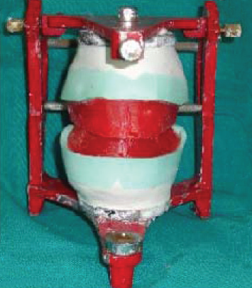
Fig 7: Jaw relation recorded and articulated.
Lower base plate construction:
An additional autopolymerising resin mandibular denture base was fabricated and a superstructure was constructed. The superstructure was made of 2 vertical self-cured pillars at established vertical dimension in the region of premolars with retentive loops made of thin orthodontic wire in between and posterior to them (Fig. 8). The superstructure had two functions: to provide even occlusal stops at the correct vertical dimension and to provide support for the neutral zone impression material. This base was placed in the mouth, checked for stability and ensured that loops and vertical pillars do not interfere with muscle movements during function (Fig. 9).

Neutral zone impression:
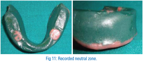
The maxillary rim was left in mouth in order to provide enough support to the facial musculature during making neutral zone impression. Then the green stick material was softened and loaded over the wire loops on buccal and lingual aspects and inserted in mouth (Fig. 10). The patient was asked to perform a series of actions designed to simulate physiological functioning which included smiling, grinning, pouting/pursing lips, count from 60 to 70, talk aloud, pronounce the vowels, sip water, swallow, slightly protrude the tongue and lick the lips. Excess compound was trimmed away and the material was resoftened and placed back into the mouth asking the patient to repeat the functional movements. The final record should be perfectly stable in place (Fig. 11).

Fig 10: Recording neutral zone with green stick compound.
Laboratory procedures:
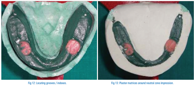
The neutral zone impression was placed over the mandibular master cast. Indexes/locating grooves were made on the mandibular cast, in order to guide the placement and removal of the plaster matrices properly (Fig. 12). Index grooves were made on side and centre of the land area of cast. Facial and lingual matrices of this denture space were made using plaster so that it is in level of occlusal plane of record and surrounds the neutral zone impression (Fig. 13). Once set, the matrices were sectioned and removed from the cast.
Then the green stick material and the adapted wire loops were removed from the mandibular record base leaving an empty space (neutral zone space) within the plaster indices. Wax was then poured into the space giving an exact representation of the neutral zone (Fig. 14). Teeth were then arranged exactly following the index. The mandibular teeth were arranged first and in accordance with the mandibular teeth, maxillary teeth were arranged. During the teeth arrangement, their position can be checked by putting the index together around the wax try-in (Fig. 15). The contours of the mandibular denture as derived from neutral zone technique were preserved and no additional wax was added on denture flanges.


Fig 15: Teeth arrangement done in neutral zone space.
Try-in and denture insertion:
Trial denture was tried in the mouth to evaluate the stability, aesthetics, phonetics and occlusion. Once the wax try-ins were deemed satisfactory the dentures were processed with heat cured acrylic, finished and polished using conventional method.On final insertion the dentures were fully inspected and evaluated for stability, aesthetics and occlusion; and any minor occlusal errors were eliminated (Fig. 16). The patient was reviewed after a week and minor denture related complaints were corrected. Results were found satisfactory and patient also was satisfied with the dentures being stable.

Fig 16: Denture inserted.
SUMMARY & CONCLUSION
Providing stable mandibular dentures for patients with severely resorbed mandibular ridges is a challenge. To overcome this problem, the dentures must be fabricated with their contours harmonizing to the surrounding muscles.
This article highlights the technique to achieve an effective retention, stability and support for Atwood’s Order VI ridge deformities. By following the combination of admix impression technique for the secondary impression and the neutral zone for the construction of lower complete denture, it would be possible to economically yet effectively rehabilitate a patient with flat, atrophic, depressed, mandibular ridge thereby improving the function. The technique is relatively simple but there are increased chair time and laboratory steps.
Disclaimer from Guident
The views and opinions expressed in articles published in Guident are solely those of the authors and do not necessarily reflect the views of the editors, the editorial board or the publisher, Ivory India. Guident does not endorse any product, technique or treatment described. Clinical content is intended for qualified dental and medical professionals and is not a substitute for individual clinical judgement. Guident, its editors and the publisher accept no liability for any consequence arising from the use of information published in the journal.
How to cite this article
Dr. Manesh Lahori, Dr. Prateek Agrawal. Amalgamation of Techniques for Rehabilitation of Severely Resorped Residual Ridge. Guident. 2013 Jul;6(9). Available from: https://ivoryindia.in/articles/prosthodontics/amalgamation-of-techniques-for-rehabilitation-of-severely-resorped-residual-ridge.html