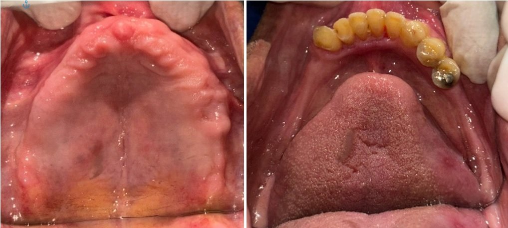
Esthetic Rehabilitation of Fluorosed Patients with Ceramic Veneers: Case Report
Department of Prosthodontics I.T.S Dental College, Ghaziabad, Muradnagar, UP, India.
ABSTRACT
Porcelain dental veneering has become a popular method of solving many aesthetical and functional dental problems. Fluorosis is endemic in distribution and occurs as a result of drinking water with high fluoride content. Although fluorosis in its mild form is not considered to be of cosmetic significance, the more severe forms affect tooth structure and can cause psychological distress to the affected individual. This paper presents two case reports with severe dental fluorosis that were managed for dental aesthetics.
Keywords:
Dental fluorosis, ceramic veneers, incisal wraparound.
INTRODUCTION
Dental fluorosis is a disturbance affecting the enamel formation related to excessive ingestion of fluoride during the time of enamel mineralization. The clinical appearance reflects a spectrum of changes. In its mild form, lustreless white lines or diffuse opacities are present, while in the more severe forms, generalized opaque and chalky appearance with confluent pitting and staining of hypomineralized tissues may be seen. Fluorotic enamel is hypomineralized and porous and, following eruption, extensive mechanical breakdown of the surface enamel and secondary staining of the underlying hard tissues will occur in a severely affected dentition.1 Therefore, severe form of fluorosis, not only disturbs enamel significantly but also affects aesthetics quite adversely and can cause psychological distress to the affected person.
During the last decade, due to growing patient demand for esthetic restorations and more conservative trends in restorative dentistry, the use of porcelain veneers has become a widespread, reliable, and successful technique for restoring discolored, worn, malformed, or fractured teeth.2 Many factors influence the long term success of porcelain veneers, such as tooth surface, porcelain thickness, type of luting agent and adhesive system, marginal adaptation, periodontal response, tooth morphology, functional and parafunctional activities, and the geometry of the preparation.2 Regarding tooth morphology, the palatal concavity and the incisal areas of maxillary anterior teeth are considered to be high stress concentration areas during tooth function3,4 this fact is physiologically compensated for by having increased enamel thickness in these zones.5 The enamel removed during tooth preparation should be compensated for with a material having enamel-like properties to restore the original biomechanical behaviour of a tooth.5 Preparation design and geometry are possible variables involved in the ultimate success of a restoration. To achieve the best adhesion, it is desirable to have at least 50% to 70% of the enamel surface available for the etching procedures.6
One of the most controversial topics regarding porcelain veneers is the geometry of the preparation.3,7,8 Seymour et al9 demonstrated a lower amount of compressive stress for the incisal overlap preparation, which the author indicated should better tolerate occlusal loading.
Typical indications for veneers include teeth that are malformed, rotated or malpositioned. Veneers may also be used to close single or multiple diastemas to change the colour of unattractive teeth, to restore abraded or eroded restorations or to cover and replace faulty restorations. Other factors such as stain etiology, occlusion and the age, health and oral hygiene of the patient are important considerations in the treatment plan.
CASE REPORT 1
A 20 years old female patient reported with chief complaint of discolored upper front teeth since past 10 years. She gave no significant medical history. The patient examination revealed generalized enamel fluorosis affecting all of the permanent teeth (Fig. 1). Confluent pitting was present on most of the surfaces of the upper teeth and only the second premolars of the lower teeth, with wide spread of yellow-brown stains.

Fig 1
Surfaces of lower anterior teeth were, however; relatively less affected than others. Oral hygiene was good. The perception of her appearance had a detrimental influence on her self- confidence; she was reluctant to smile due to appearance of her teeth. At the same time, she was afraid to alter her teeth, and wanted a conservative treatment alternative. She was given the option of composite or ceramic veneers. The patient decided to go in for ceramic veneers. The treatment plan accepted by the patient was constructing 06 porcelain laminate veneers (PLV) on the upper teeth # 13 to # 23.
Tooth preparation
The following technique was used. The preparation was conservative, but the space for porcelain should be of at least 0.5 mm. After marking the facial preparation, with indelible pen, we used a depth cutting diamond for providing the proper depth reduction.
Incisal reduction is an important factor in the long-term fracture resistance of veneers. The incisal edge was reduced by 1.5 mm, and a lingual chamfer was prepared. This chamfer exposes porcelain to compression instead of shearing, during the initial phase of protrusive movement, and as long as forces are against the tooth, fracture resistance is high.
The facial reduction was 0.75 mm incisal and 0.5 mm cervical, with a chamfer finish line in a slightly supragingival location [Fig 2]. After displacement of gingival tissues with a retraction cord, impression was made with polyvinyl siloxane. Provisional restorations from A2 shade composite resin were prepared.
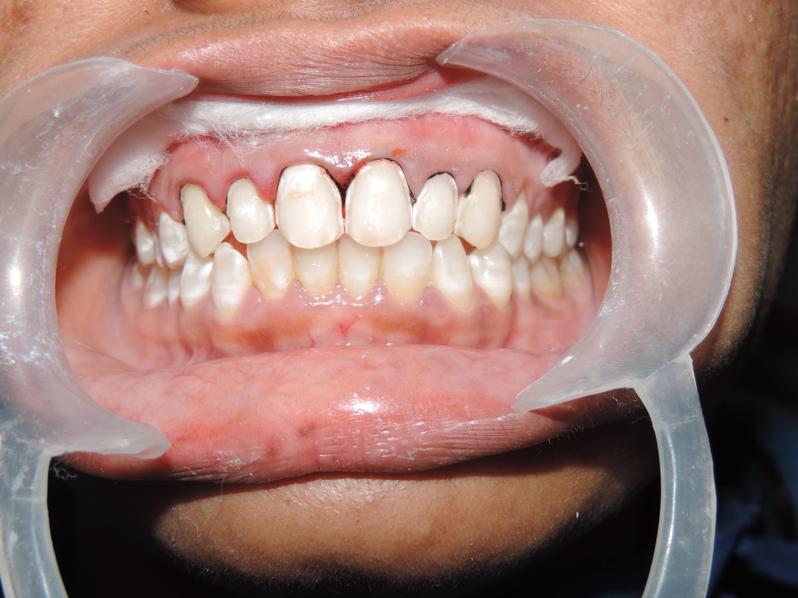
Fig 2
Laboratory phase
The laminate veneers were made from IPS Empress using the lost wax technique [Fig 3,4]. The flexural strength of this type of ceramics is well documented. When returned from the laboratory, the veneers were tried for fit and shade matching.
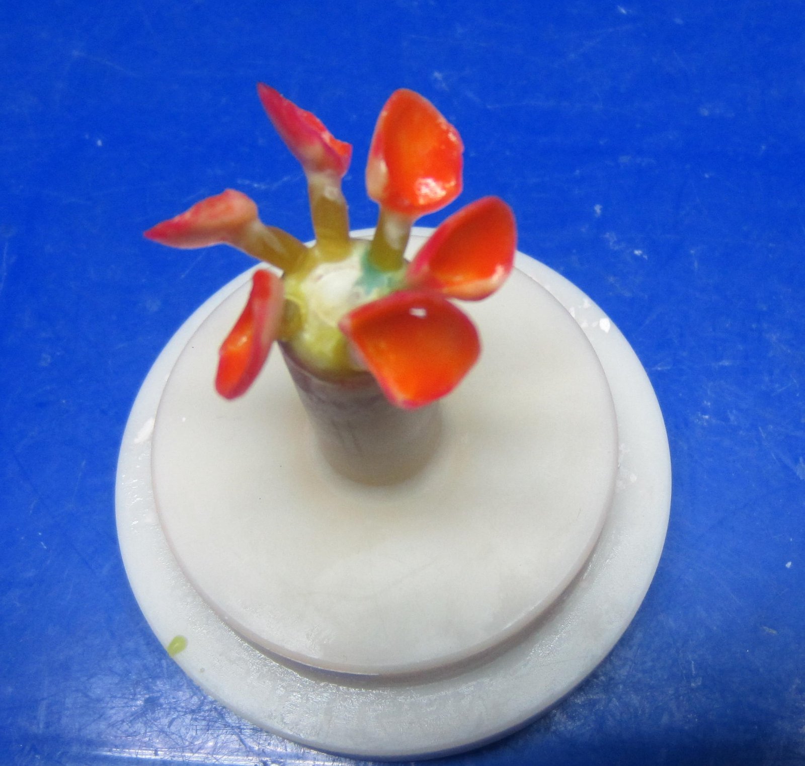
Fig 3
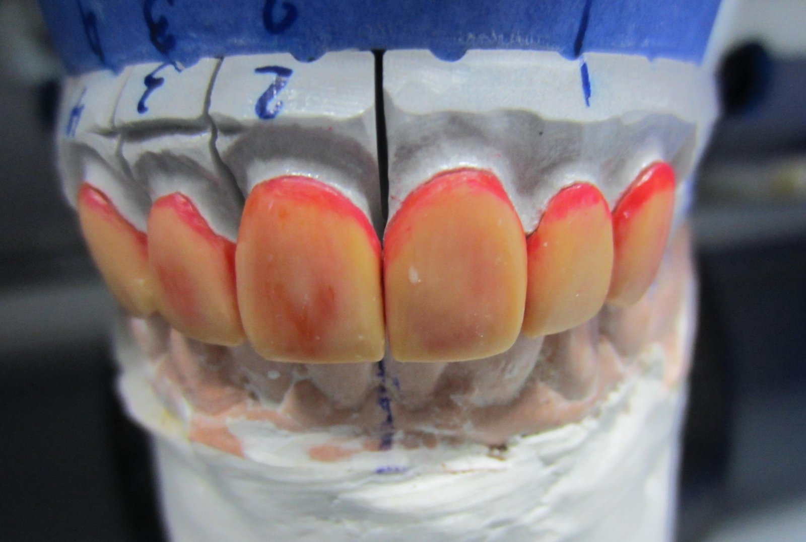
Fig 4
Cementation
For cementation of the ceramic veneers, resin luting agent was used because it provides superior esthetics, strength and optimal wear resistance. The cementation consisted of several steps: etching the veneers with hydrofluoric acid, applying the silane coupling agent, preparing the teeth (isolation, etching, and bonding), preparing and applying the resin cement, and light-curing. After finishing and polishing, the patient was satisfied with the final esthetic result [Fig 5,6].
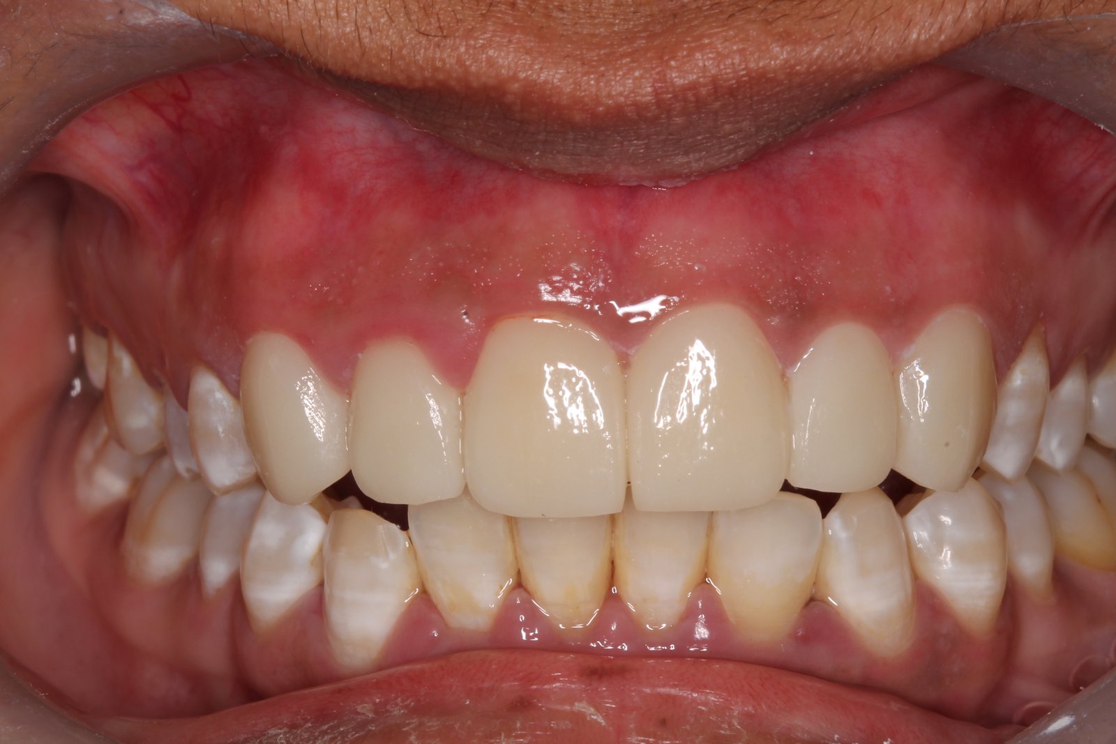
Fig 5
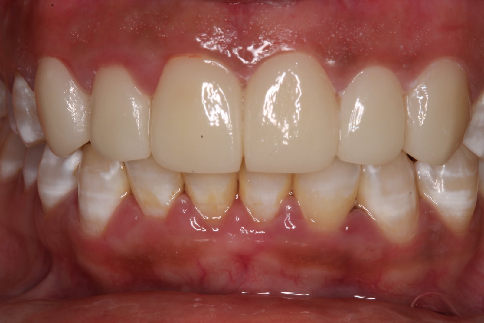
Fig 6
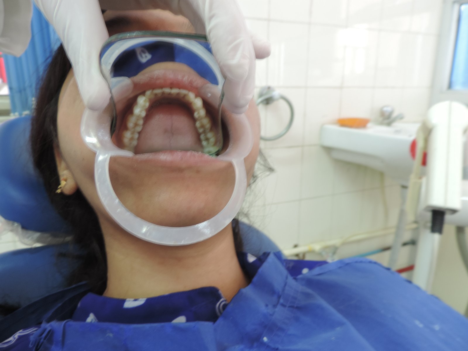
Figure-7: Occlusal view mandible
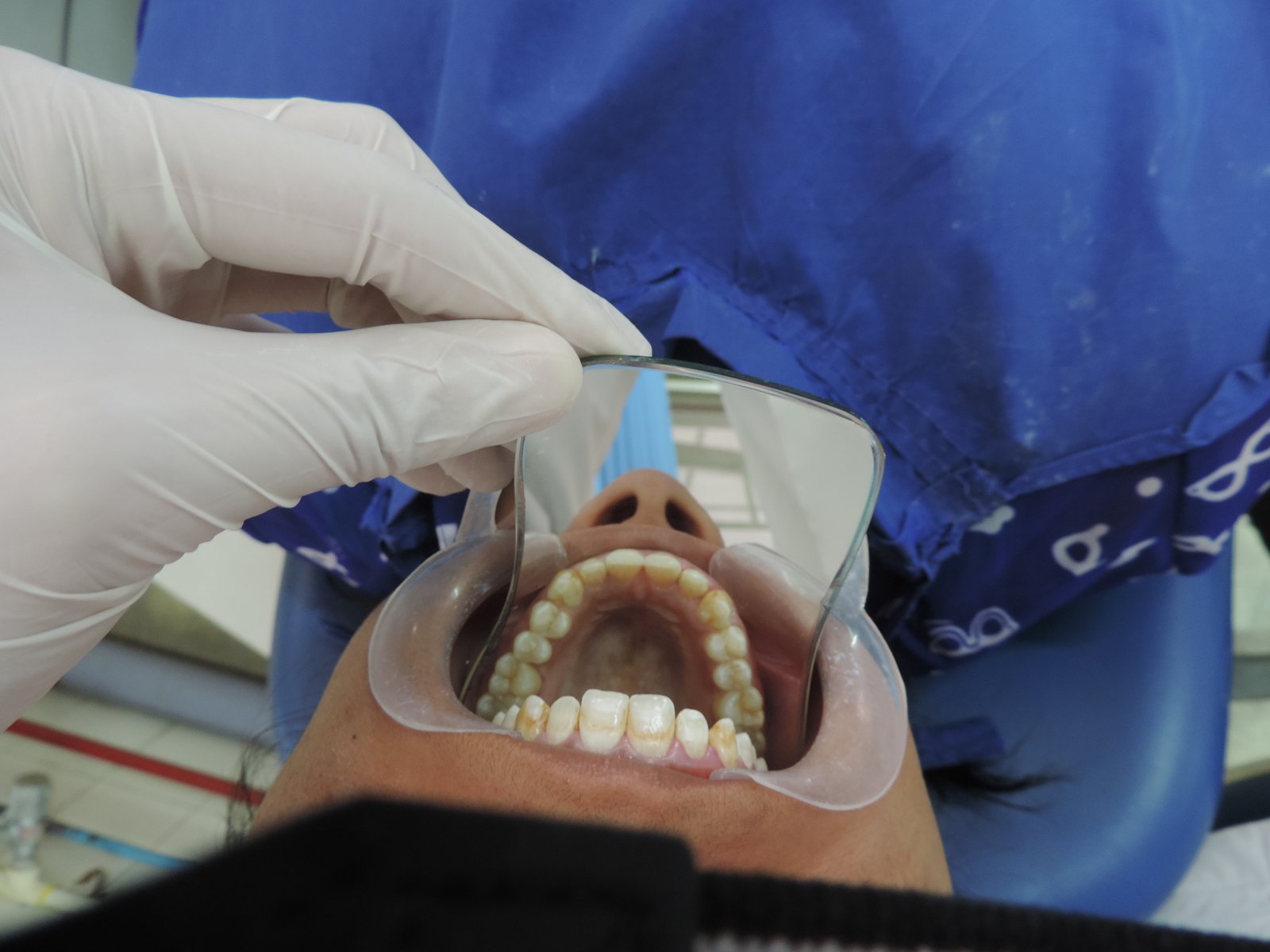
Figure-8: Occlusal view maxillary
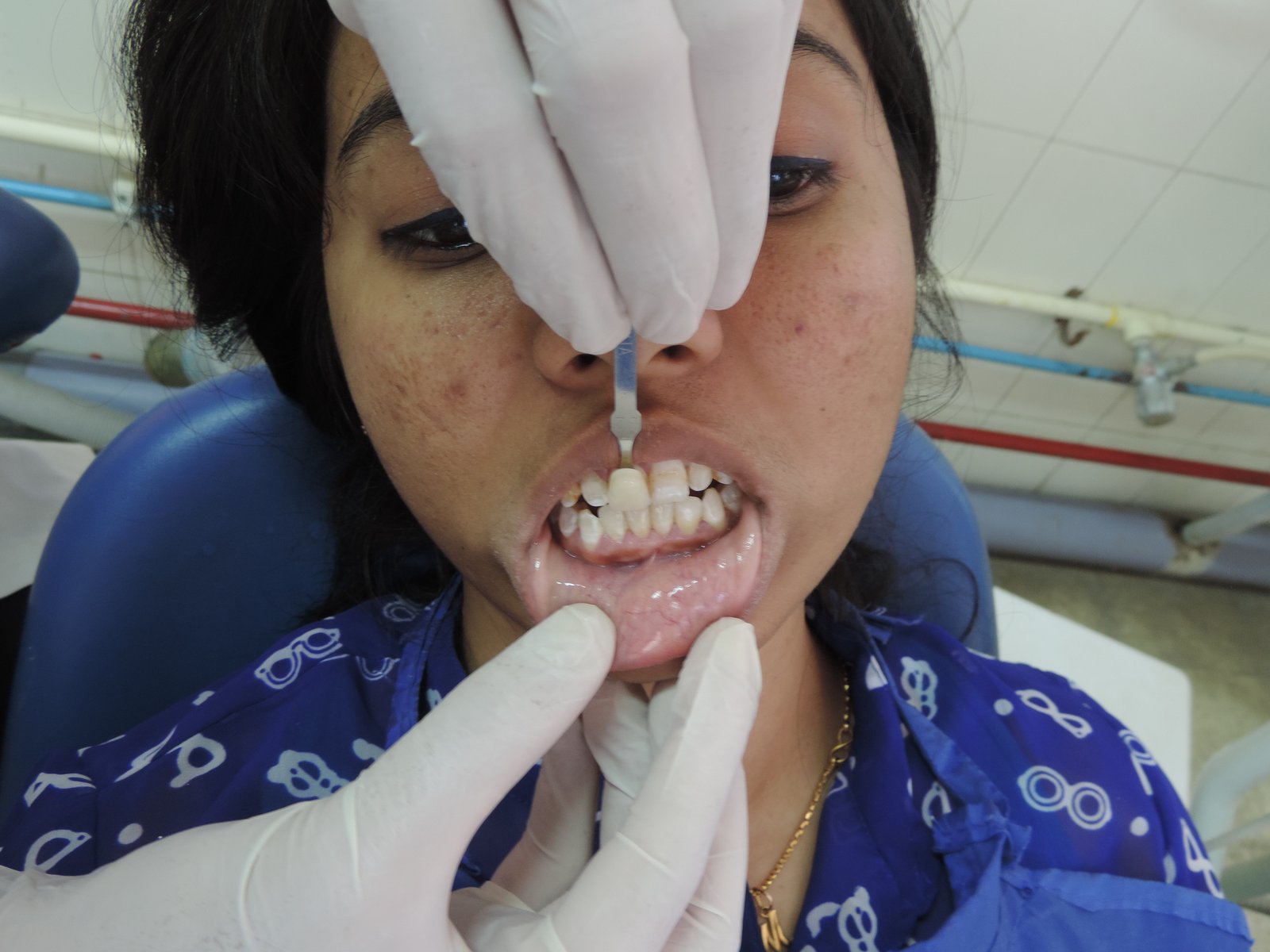
Figure-9: Shade selection
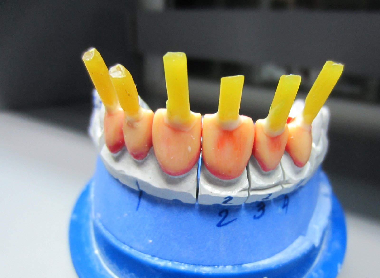
Figure 10: wax pattern
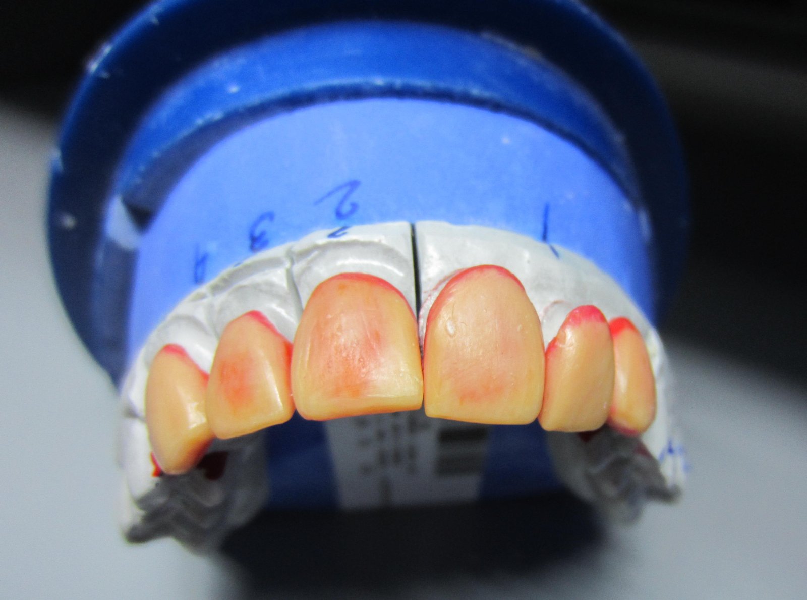
Figure 11: maxillary labial surface wax pattern
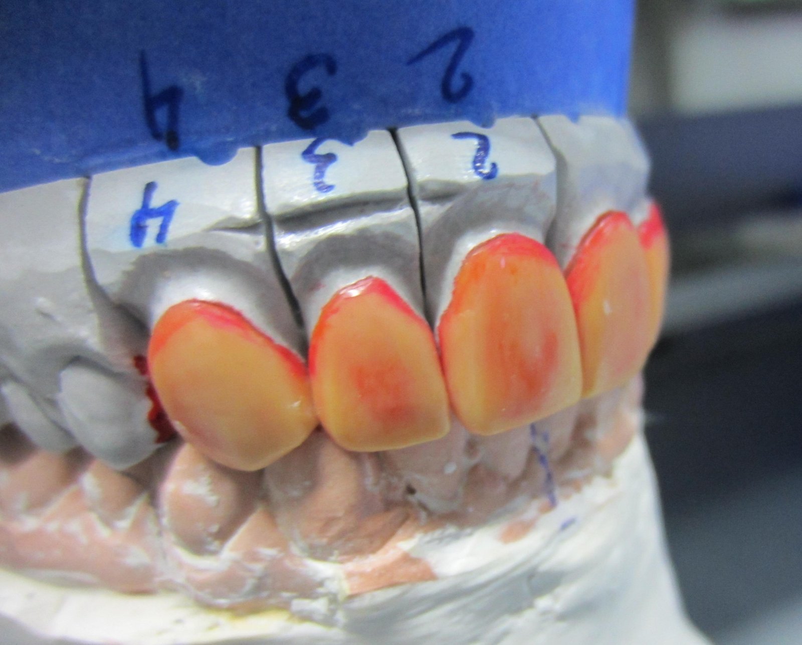
Figure 12: right wax pattern view
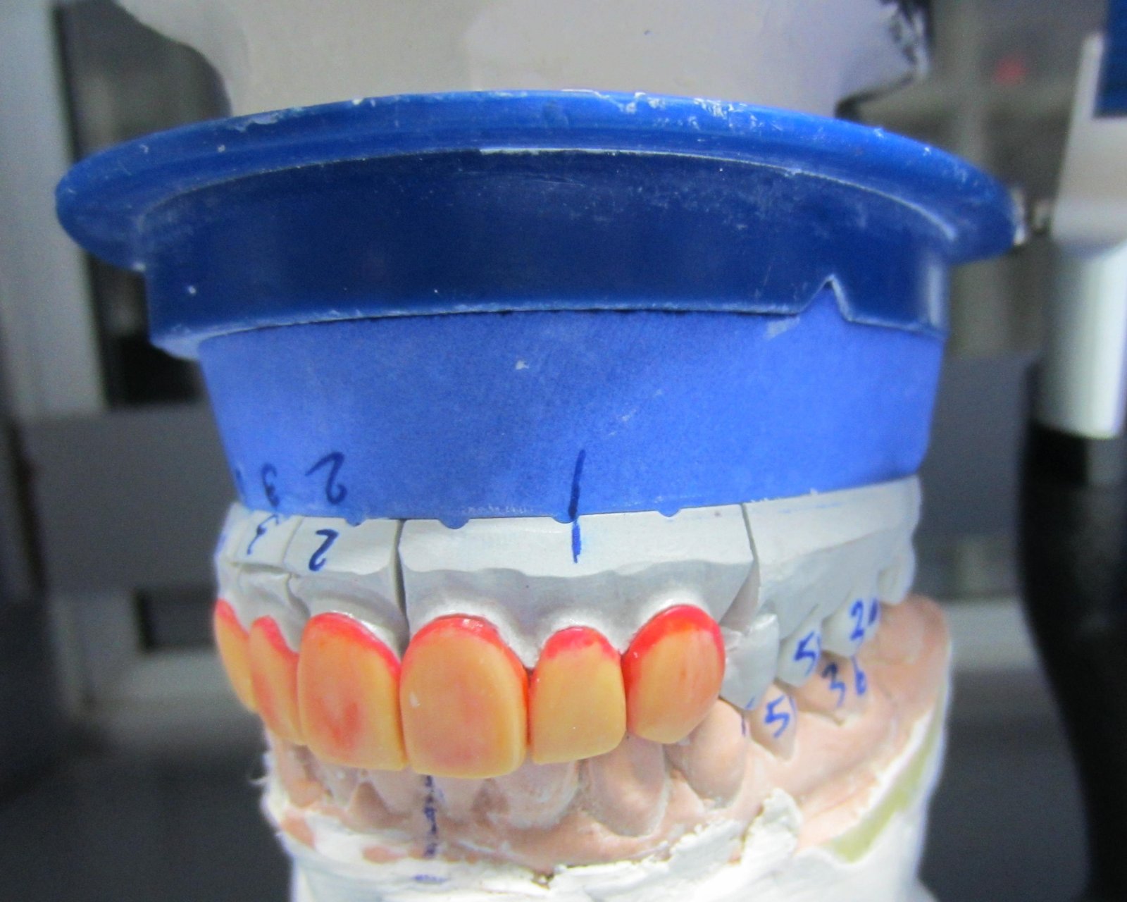
Figure 13: left wax pattern view
DISCUSSION
Clinical examination revealed discolored enamel, confluent pitting and corrugated incisal edges. Bleaching or microabrasion of severely fluorosed teeth is often ineffective or gives transient results, while composite resin laminates veneers not only discolor and wear with time, but quite often become chipped or debonded. Small localized intrinsic discolorations or defects that are surrounded by healthy enamel are ideally treated with direct partial veneers (i.e composites). Since the treatment plan warrants incisal defect restoration and lengthening of cervicoincisal length, incisal “wraparound” design was decided for biomechanical preparation. Additionally, the incisal lapping design allows for improved esthetics along the incisal edge and accurate seating of the laminates.Ceramic laminate veneers are more hygienic and have good lustre over composites. The esthetics are considerably better than any other veneering material because of the ability to control color and surface texture with ceramics. They can be stained both internally and superficially and have a natural fluorescence, lending a certain vitality.
In both the cases IPS Empress ceramic laminate system was selected over composites, CAD/CAM laminates and those fabricated by refractory investment layer-on technique. The CAD/CAM laminate system uses conventional cements, hence lacks retention. Moreover, the biomechanical preparation is not conservative and the laminates are more opaque. Laminates prepared from the layer-on technique have low marginal strength compared to pressable ceramics. Because they are more translucent, any mismatch between shades of luting cement and laminates might compromise the final esthetic outcome.
The flexural strength of the IPS Empress leucite glass ceramic has been improved to 160 ± 8 MPa. Laminates fabricated using the lost-wax technique feature a particularly high accuracy of fit, as there is no sinter shrinkage. The tooth shade is determined on the cleaned, unprepared tooth or adjacent tooth with the help of the Ivoclar Vivadent Chromascop shade guide provided by the manufacturer. Hence it provides more life- like appearance. IPS Empress Esthetic restorations have to be adhesively cemented. Variolink II Try-In pastes are water-soluble that allow the shade of the ceramic restoration to be simulated prior to final cementation. The Variolink II Try-In pastes feature the same optical properties as the subsequently used luting composite Variolink II. Therefore, the system provides several advantages over other laminate systems.
Porcelain dental veneers are not the only alternative for all esthetic abnormalities, they are truly a remarkable restoration when they are the treatment of choice. As glazed porcelain retains its color and is wear resistant and biocompatible, laminate veneers made from this material can be the restoration of choice for severely fluorosed teeth.
REFERENCES
- Thylstrup A and Fejerskov O. Clinical appearance of dental fluorosis in permanent teeth in relation to histopathologic changes. Community Dent Oral Epidemiol 1978; 6:315-28.
- Peumans M, Van Meerbeek B, Lambrechts P, Vanherle G. Porcelain veneers: a review of the literature. J Dent 2000;28:163-77.
- Highton R, Caputo AA, Matyas J. A photoelastic study of stresses on porcelain laminate preparations. J Prosthet Dent 1987;58:157-61.
- Nicholls JI. Tensile bond of resin cements to porcelain veneers. J Prosthet Dent 1988;60:443-7.
- Magne P, Oh WS, Pintado MR, DeLong R. Wear of enamel and veneering ceramics after laboratory and chairside finishing procedures. J Prosthet Dent 1999;82:669-79.
- Chiche GJ, Pinault A. Esthetics of anterior fixed prosthodontics. Chicago:Quintessence; 1994. p. 62-4.
- Meijering AC, Creugers NH, Roeters FJ, Mulder J. Survival of three types of veneer restorations in a clinical trial: a 2.5-year interim evaluation. J Dent 1998;26:563-8.
- Hui KK, Williams B, Davis EH, Holt RD. A comparative assessment of the strengths of porcelain veneers for incisor teeth dependent on their design characteristics. Br Dent J 1991;171:51-55
- Seymour KG, Cherukara GP, Samarawickrama DY. Stresses within porcelain veneers and the composite lute using different preparation designs. J Prosthodont 2001;10: 16-21.
Disclaimer from Guident
The views and opinions expressed in articles published in Guident are solely those of the authors and do not necessarily reflect the views of the editors, the editorial board or the publisher, Ivory India. Guident does not endorse any product, technique or treatment described. Clinical content is intended for qualified dental and medical professionals and is not a substitute for individual clinical judgement. Guident, its editors and the publisher accept no liability for any consequence arising from the use of information published in the journal.
How to cite this article
Dr. Sunil Pal, Dr. Arjita Dutta, Dr. Pankaj Dutta. Esthetic Rehabilitation of Fluorosed Patients with Ceramic Veneers: Case Report. Guident. 2025 Jul;18(8). Available from: https://ivoryindia.in/articles/prosthodontics/esthetic-rehabilitation-of-fluorosed-patients-with-ceramic-veneers-case-report.html