Esthetic Rehabilitation Using Anatomic Post and Lithium Disilicate Crownintroduction: A Case Report
Department of Prosthodontics, Government Dental College & Hospital, Chhatrapati Sambhajinagar
ABSTRACT
Restoration of endodontically treated anterior teeth with flared root canals presents both functional and esthetic challenges due to the loss of substantial coronal and radicular dentin. This case report describes the esthetic and structural rehabilitation of a severely compromised maxillary anterior tooth using a customized anatomic fiber post and a lithium disilicate ceramic crown. An anatomic post was fabricated by relining a prefabricated fiber post with composite resin directly within the root canal to achieve optimal adaptation and stress distribution. The coronal portion was restored using an adhesively bonded lithium disilicate crown to achieve high esthetic integration and strength. This conservative approach successfully restored the form, function, and esthetics of the tooth while preserving remaining tooth structure. The case highlights the clinical advantages of using anatomic posts and modern adhesive restorative materials in managing flared canals with minimally invasive techniques.
INTRODUCTION
The restoration of endodontically treated teeth, particularly those in the anterior region, poses both functional and esthetic challenges, especially when significant loss of coronal and radicular tooth structure is present.1 In cases involving flared or over-enlarged root canals, traditional prefabricated fiber posts often fail to achieve adequate adaptation to the canal walls, resulting in thick layers of resin cement, increased polymerization shrinkage, and compromised stress distribution.2,3 This mismatch can jeopardize the long-term prognosis of the restoration due to potential debonding or root fracture.4
To address these limitations, the concept of the anatomic or customized fiber post has been introduced.5 This technique involves relined or customized adaptation of a fiber post using a resin composite material that precisely conforms to the internal anatomy of the flared root canal.6 This approach offers several advantages: it reduces the volume of resin cement needed, improves post retention, enhances load distribution along the root, and reinforces weakened root structures. Moreover, it preserves the biomechanical integrity of the tooth, minimizing the risk of catastrophic failure.2,7
From an esthetic standpoint, the anterior zone demands materials that not only restore function but also mimic the translucency and optical properties of natural dentition.8 Lithium disilicate crowns are among the most preferred choices for restoring anterior teeth due to their excellent esthetics, superior strength, biocompatibility, and adhesive bonding capabilities.9,10 When used in combination with a well-adapted fiber post, these restorations can offer highly satisfactory clinical outcomes in terms of both appearance and durability.11
This case report describes the interdisciplinary management of a structurally compromised, flared maxillary anterior tooth using a customized anatomic fiber post technique, followed by esthetic rehabilitation with a lithium disilicate crown. The report highlights the clinical rationale, procedural details, and advantages of this conservative yet effective approach, emphasizing the importance of individualized post adaptation in achieving long-term restorative success
CASE REPORT
23 year old female patient reported to department of prosthodontics having chief complaint of frequent dislodgement prosthesis in upper front teeth region. Dental history revealed that patient had undergone root canal treatment and crown prosthesis 4-5 years back with 21. On clinical examination 3-4 mm tooth structure. Radiographic examination revealed a wide and flared canal. The treatment plan included customized anatomic post fabrication and full-coverage esthetic crown restoration using lithium disilicate ceramic.
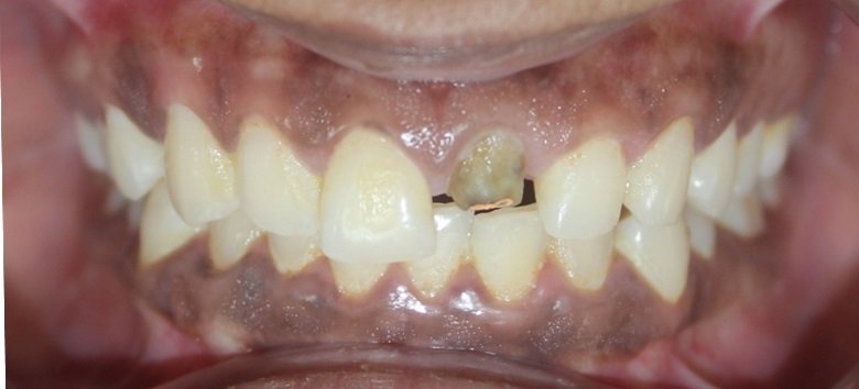
Fig 1: Preoperative photograph
PROCEDURE
Canal Preparation:
The existing root canal filling was evaluated and confirmed to be intact. The post space was prepared by removing gutta-percha using PEESO reamers (MANI, Japan) while maintaining a 4–5 mm apical seal.
Fig 2: Fabrication of fiber post;
- A: Acid etching of fiber post
- B: Application of bonding agent
- C: Composite resin adapted over fiber post
- D: Anatomic post snugly fitting in post
- E: Anatomic post fabricated
- F: Curing was done extroral


Fig 3: Anatomic post core cemented and core build followed by Tooth Preparation and gingival retraction
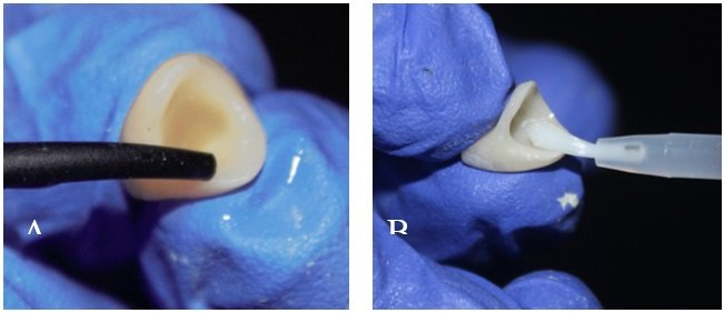
Fig 4: Surface treatment for lithium disilicate crown- A: Application of Hydroflouric Acid followed by water rinsing, B: Application a ceramic primer
- Post Customization:
A prefabricated translucent glass fiber post (Mailyard fiber post) of size 4 was chosen and assessed for fit, revealing a poor fit within the canal. Decision was made to construct an anatomical post with relining. The canals were dried and coated with petroleum jelly. Silane (Ultradent, USA) was applied to the intended fiber posts for one minute. Selected fiber post was relined with flowable composite resin (3M's Filtek Supreme Flowable) inside the lubricated canal to mimic the canal anatomy. The relined post was then initial light curing for 20 seconds from above followed by light-cured outside the canal. The prepared anatomic post was then re-checked intraorally for proper fit, retention and adaptation, demonstrating a snug fit and secure retention in the post space. - Post Cementation:
The canal was cleaned and etched. A dual cure resin cement was used to bond the customized anatomic post within the canal. Excess cement was removed, and the area was light-cured. - Core Build-up:
A core build-up was performed using nanohybrid composite (Prevest Fusion Universal Composite) resin to restore the lost coronal structure and provide adequate support for the final crown. Tooth Preparation and Crown Fabrication:
The tooth was prepared for an all-ceramic crown. Gingival retraction was achieved using Sure-endo 000 retraction cord, and a silicone impression (Dentsply Aquasil impression material) was created. A CAD-CAM lithium dislicate crown was fabricated and cemented using dual-cure resin cement.
Fig 5: Final lithium disilicate crown cemented with 21 under rubber dam isolation
- A: Tooth surface is cleaned with a polishing brush and fluoride-free cleaning paste
- B: Application of phosphoric acid
- C:Rinsing thoroughly with water
- D: Application of bonding agent
- E: Cementation of crown and Light curing was done
- F: Excess cement was removed with floss


Fig 6: Post- operative photograph
Cementation:
The lithium disilicate crown was tried in, checked for fit, esthetics, and occlusion. Cementation was carried out with resin cement (Variolink N Clear )according to manufacturer instruction using resin cement.
Periodic follow-ups were conducted.
DISCUSSION
The rehabilitation of endodontically treated teeth, especially those with flared or excessively tapered canals, presents both mechanical and esthetic challenges.2 Traditional prefabricated posts often fail to adapt adequately to wide or irregular canal spaces, resulting in a thick layer of luting cement that can compromise retention and increase the risk of post debonding and root fracture.8 In such cases, the use of an anatomic fiber post, which is customized to the internal geometry of the root canal, offers a significant clinical advantage.12,13
Anatomic posts are created by relining prefabricated fiber posts with composite resin directly within the prepared canal space. This technique improves post adaptation, reduces the volume of luting cement, and enhances the post-to-dentin bond strength. Moreover, fiber posts have a modulus of elasticity similar to that of dentin, which contributes to a more favorable stress distribution along the root, reducing the incidence of catastrophic failures compared to rigid metallic or ceramic posts.
In the presented case, the use of a customized anatomic post allowed for conservative restoration of a structurally compromised anterior tooth with thin root walls. This technique avoided the need for extensive canal modification or use of multiple posts, thereby preserving the remaining tooth structure. Additionally, it ensured intimate adaptation of the post to the canal anatomy, which is critical in achieving long-term success in teeth with flared canals.
The coronal portion of the tooth was restored using a lithium disilicate ceramic crown, a material that combines excellent esthetics, translucency, and flexural strength. Lithium disilicate (e.g., IPS e.max) is particularly well-suited for anterior teeth due to its ability to mimic natural enamel while maintaining functional durability. When adhesively bonded, it forms a monoblock with the underlying composite core and post system, further reinforcing the restored tooth.
Adhesive cementation plays a vital role in the success of both the post and crown. In this case, the post was luted using a dual-cure resin cement to ensure optimal bonding even in areas with limited light access. Similarly, the lithium disilicate crown was bonded using an esthetic resin cement, providing a seamless blend with adjacent teeth and minimizing marginal leakage.
This case highlights the importance of minimally invasive, biomimetic restorative approaches in the management of endodontically treated teeth. By utilizing materials with mechanical properties that closely resemble natural tooth structure, and by customizing the restoration to the individual anatomy of the tooth, clinicians can achieve predictable outcomes that restore both esthetics and function.
Furthermore, follow-up evaluation showed no signs of mobility, discoloration, or periapical pathology, indicating the long-term viability of this approach. The successful outcome reinforces the use of anatomic posts and adhesive ceramic restorations as a conservative and esthetically superior alternative for complex anterior tooth rehabilitation.
CONCLUSION
The restoration of endodontically treated teeth with flared canals requires a careful balance of biomechanics, adhesion, and esthetics. This case highlights the effectiveness of using a customized anatomic fiber post to achieve optimal adaptation within a compromised canal, thereby enhancing retention and stress distribution. The use of a lithium disilicate ceramic crown provided excellent esthetic integration and long-term durability. Together, this adhesive and minimally invasive approach offers a predictable and conservative solution for the functional and esthetic rehabilitation of structurally compromised anterior teeth. Continued clinical follow-up is essential to evaluate long-term success.
REFERENCES
- Lee JH, Sohn DS, Lee CH. Fabricating a fiber-reinforced post and zirconia core with CAD/CAM technology. J Prosthet Dent. 2014 Sep;112(3):683-5. doi: 10.1016/j.prosdent.2014.01.015. Epub 2014 Mar 27. PMID: 24680166.
- Santos Pantaleón D, Tribst JPM, García-Godoy F. Influence of size-anatomy of the maxillary central incisor on the biomechanical performance of post-and-core restoration with different ferrule heights. J Adv Prosthodont. 2024 Apr;16(2):77-90. doi: 10.4047/jap.2024.16.2.77. Epub 2024 Apr 24. PMID: 38694191; PMCID: PMC11058349.
- Katkade AS, Rajguru VL, Mahale KM, Khalikar SA, Mahajan SV, Tandale U. An innovative dental technique to manage wide & Flared root canals using an anatomical intra-radicular retainer. International Dental Journal of Student’s Research 2024;12(1):50-54.
- Libonati A, Di Taranto V, Gallusi G, Montemurro E, Campanella V. CAD/CAM Customized Glass Fiber Post and Core With Digital Intraoral Impression: A Case Report. Clin Cosmet Investig Dent. 2020 Feb 10;12:17-24. doi: 10.2147/CCIDE.S237442. PMID: 32104100; PMCID: PMC7021168.
- Signore A, Kaitsas V, Ravera G, Angiero F, Benedicenti S. Clinical evaluation of an oval-shaped prefabricated glass fiber post in endodontically treated premolars presenting an oval root canal cross-section: a retrospective cohort study. Int J Prosthodont. 2011 May-Jun;24(3):255-63. PMID: 21519574.
- Shetty A, Raghu R, Shetty A, Gautham PM. Anatomic post and core- A novel approach to restore flared root canals: A case report. Int J Oral Health Dent 2019;5(1):41-43.
- Bitter K, Kielbassa AM. Post-endodontic restorations with adhesively luted fiber-reinforced composite post systems: a review. Am J Dent. 2007 Dec;20(6):353-60. PMID: 18269124.
- Grandini S, Sapio S, Simonetti M. Use of anatomic post and core for reconstructing an endodontically treated tooth: a case report. J Adhes Dent. 2003 Fall;5(3):243-7. PMID: 14621246.
- Leandrin TP, Fernández E, Lima RO, Besegato JF, Escalante-Otárola WG, Kuga MC. Customized Fiber Post Improves the Bond Strength and Dentinal Penetrability of Resin Cementation System to Root Dentin. Oper Dent. 2022 Jan 1;47(1):E22-E34. doi: 10.2341/20-117-L. PMID: 34969119.
- Iglesia-Puig MA, Arellano-Cabornero A. Fiber-reinforced post and core adapted to a previous metal ceramic crown. J Prosthet Dent. 2004 Feb;91(2):191-4. doi: 10.1016/j.prosdent.2003.11.004. PMID: 14970767.
- Veríssimo C, Simamoto Júnior PC, Soares CJ, Noritomi PY, Santos-Filho PC. Effect of the crown, post, and remaining coronal dentin on the biomechanical behavior of endodontically treated maxillary central incisors. J Prosthet Dent. 2014 Mar;111(3):234-46. doi: 10.1016/j.prosdent.2013.07.006. Epub 2013 Nov 5. PMID: 24199605.
- Sary S B, Samah M S, Walid A AZ. Effect of restoration technique on resistance to fracture of endodontically treated anterior teeth with flared root canals. J Biomed Res. 2019 Apr 22;33(2):131-138. doi: 10.7555/JBR.32.20170099. PMID: 30418168; PMCID: PMC6477170.
- Alshetiwi DSD, Muttlib NAA, El-Damanhoury HM, Alawi R, Rahman NA, Elsahn NA, Karobari MI. Evaluation of mechanical properties of anatomically customized fiber posts using E-glass short fiber-reinforced composite to restore weakened endodontically treated premolars. BMC Oral Health. 2024 Mar 11;24(1):323. doi: 10.1186/s12903-024-04102-2. PMID: 38468269; PMCID: PMC10926594.
Disclaimer from Guident
The views and opinions expressed in articles published in Guident are solely those of the authors and do not necessarily reflect the views of the editors, the editorial board or the publisher, Ivory India. Guident does not endorse any product, technique or treatment described. Clinical content is intended for qualified dental and medical professionals and is not a substitute for individual clinical judgement. Guident, its editors and the publisher accept no liability for any consequence arising from the use of information published in the journal.
How to cite this article
Dr. Aishwarya Gholap, Dr. Kishor Mahale, Dr. Smita Khalikar. Esthetic Rehabilitation Using Anatomic Post and Lithium Disilicate Crownintroduction: A Case Report. Guident. 2025 Aug;18(9). Available from: https://ivoryindia.in/articles/prosthodontics/esthetic-rehabilitation-using-anatomic-post-and-lithium-disilicate-crownintroduction-a-case-report.html