Occlusal Concepts in Full Mouth Rehabilitation-a Review
Department of Prosthodontics and Crown & Bridge, Shree Bankey Bihari Dental College and Research Centre, Ghaziabad Dr. Yashi Garg (Senior Lecturer), Department of Prosthodontics and Crown & Bridge, Inderprastha Dental College and Hospital, Ghaziabad UP.
INTRODUCTION
Complete rehabilitation of the dentition is a complex, integral part of prosthetic dentistry, requiring knowledge of physiology, anatomy, histology and physics, as well as a thorough understanding of, and ability to perform, the operative and restorative phases involved. The indications of occlusal rehabilitation involve the restoration of multiple teeth which are missing, worn, broken down or decayed, to replace improperly designed and executed crown and bridge framework and treatment of temporomandibular disorders (Lucia, 1983). The objective of occlusal rehabilitation is to re-establish a state of functional as well as biological efficiency where teeth and their periodontal structures, the muscles of mastication, and the temporomandibular joint (TMJ) mechanisms all function together in synchronous harmony (Kazis et al., 1960). Proper evaluation followed by definitive diagnosis is mandatory as the aetiology of severe occlusal tooth wear is multifactorial and variable. Careful assessment of the patient’s diet, eating habits along with the present state of occlusion is essential for appropriate treatment planning (Lerner, 2008).
PHILOSOPHIES OF OCCLUSAL SCHEMES
The ideal occlusion for eccentric movements can be classified by three schemes according to the tooth contact condition; mutually protected articulation, group function, and balanced articulation. The balanced occlusion concept is applied to complete denture patients while mutually protected occlusion and group function are applied for natural dentition. The article overviews the various occlusal concepts to help absolve the complexities related to treatment planning and rehabilitation of patients requiring full mouth reconstruction.
GNATHOLOGICAL SCHEME (Pokorny et al., 2008)
Stallard was the first to coin the term gnathology in year 1924 and defined it as the science that relates to the anatomy, histophysiology and pathology of the stomatognathic system and that includes treatment of this system on the basis of examination, diagnosis and treatment planning. An early concept of comprehensive dentistry originated from the gnathologic society founded by McCollum in 1926. McCollum together with Stuart published their classic ‘‘Research Report’’ in 1955 and gave the Gnathological Concept (Stuart, 1981).
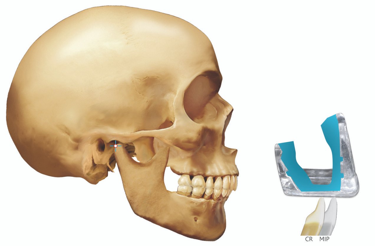
According to this scheme the movements of the condyles in the fossae determine the occlusal form. To accomplish the occlusal rehabilitation according to this scheme a fully adjustable instrument is required. The There should be simultaneous interocclusal contacts of all posterior teeth in CRCP (centric relation contact position), with forces directed axially over the long axis. In this, CRCP and IP (intercuspal position) may coincide. The closure in CRCP is followed by squeezing of the jaws together and there should not be any perceptible displacement or ‘slide’ of the mandible.
In any excursive or protrusive movement, anterior or canine guidance should separate (disocclude) the posterior teeth. If anterior guidance cannot be provided, then guidance should be placed as far forwards as possible. In this scheme, the anterior teeth contact lightly in CRCP. The palatal concavities of the anterior teeth are determined by condylar guidance (Pokorny et al., 2008). The case is waxed on a fully adjustable articulator and cusp-fossae-tripod contacts are provided (Stuart et al., 1981).
FREEDOM IN CENTRIC CONCEPT (Schuyler, 1953)
In this concept, ‘‘there is a flat area in the central fossae upon which opposing cusps contact which permits a degree of freedom (0.5–1 mm) in eccentric movements uninfluenced by tooth inclines’’. It relies on cusp-to-surface mechanics. It was suggested that incisal guidance without freedom of movement from a centric relation occlusion to a more anterior tooth intercuspation will ‘‘lock-in’’ the posterior occlusion (Schuyler, 1953). The term ‘long centric’ for freedom in centric was first used by Dawson. Long centric accommodated changes in head position and postural closure. The measurable amount of long centric needed is the difference between centric related closure and postural closure which is rarely more than 0.5 mm (Dawson, 1989).
In this scheme, cusp tip to fossa occlusion must be provided as this relation is easier to provide than tripod contact but it results in accelerated wear of occlusal surface of teeth. These areas of wear can occur between cusps and fossae, leading to a loss of support cusp contacts and possible instability. Although advocated as a simple technique, careful determination of mandibular movements is necessary for accuracy (Schuyler, 1959).
PANKEY–MANN–SCHUYLER PHILOSOPHY OF COMPLETE OCCLUSAL REHABILITATION?
This philosophy was introduced by Pankey, utilizing the principles of occlusion advocated by Schuyler, known as the Pankey–Mann–Schuyler (PMS) Philosophy of Oral Rehabilitation (Dawson, 1989) (Schuyler, 1953) (Pankey & Mann, 1960). Their philosophy was pertinently based on the spherical theory of occlusion, the ‘‘wax chew-in’’ technique and on the importance of cuspid teeth (Meyer, 1938) (Brenner, 1940) (D’Amico, 1961). As a modification of canine disclusion, this philosophy was to have group function (simultaneous contacts of the canine and posterior teeth in the working excursion) and only anterior teeth contact in the protrusive excursive movement (Mann and Pankey, 1963).
In this scheme, the PM instrument was used to establish functional occlusal plane on the mandibular teeth (Mann and Pankey, 1963). The ‘‘wax chew-in’’ technique was modified and the occlusal scheme was developed by intraoral recording of the functional occlusal path (Meyer, 1938) (Brenner, 1940). In this, both maxillary cuspids had to be in good functional contact in centric and eccentric positions before beginning the reconstruction of the posterior teeth. If not, it must be obtained by reconstruction of the cuspids even if there is no caries.
In PMS technique, the incisal guidance was the developed intraorally with acrylic resin to satisfy esthetic and functional requirements. Optimal occlusal plane is selected as dictated by the curve of Monson and mandibular posterior teeth are restored in harmony with the anterior guidance such that they will not interfere with the condylar guidance. Maxillary posterior occlusal surfaces are developed after the completion of mandibular restorations by the functionally generated path technique (FGP) (Meyer, 1938). The definitive restorations are equilibrated into a centric relation position with mandibular buccal cusps onto a flattened fossa–marginal ridge contact, with ‘‘long centric’’ incisal guidance and group function in working excursion. Use of FGP records allows eliminating all occlusal interferences and establishing functional form of the occlusal surfaces of the restoration. The PM philosophy was developed and its use advocated on a non-arcon articulator, which may not accept interocclusal records made at increased OVD (Shillingburg et al., 1997).
The advantages of this scheme is that, it is possible to plan treatment for the entire rehabilitation before a single tooth is prepared. It is a well recognized, logical procedure that progresses smoothly with less wear and tear of the dentition. There is never a need for preparing more than 8 teeth at a time. It divides the rehabilitation into several series of appointments. There is no danger of getting “lost at sea” and losing the patient’s present vertical dimension. The functionally generated path and centric relation are taken on the occlusal surface of teeth to be re-build at the exact vertical dimension to which the case will be constructed. This procedure is not time consuming and is simple also laboratory procedures are controlled to an extremely fine degree by the dentist (Dawson, 1974).
HOBO’S TWIN STAGE PROCEDURE
According to this scheme it is revealed that anterior guidance influenced the working condylar path and concluded that they were dependent factors. The posterior disclusion is dependent on; the angle of hinge rotation created by the angular difference between anterior guidance and condylar path and on inclination and shape of posterior cusps which helps in controlling harmful lateral forces.
In this technique, molar disclusion is achieved by the use of two incisal tables. The first incisal guide table termed as the incisal table without disclusion is used to fabricate restorations for posterior teeth. The second incisal table termed as the incisal table with disclusion is used to achieve incisal guidance with posterior disclusion (Hobo, 1991).
The cusp angle is considered as the most reliable determinant of occlusion as cusp angle does not deviate and is 4 times more reliable than the condylar and incisal path which show deviation (Hobo and Takayama, 1997) (Sekikawa et al., 1983). Though independent of condylar path as well as incisal path, a standard value for cusp angle was determined such that it may compensate for wear of natural dentition due to caries, abrasion and restorative works. By using the standard cusp angle, it was possible to establish the standard amount of disclusion (see Table 1). Different adjustment values of an articulator were determined for each occlusal scheme to reproduce the standard amount of disclusion. Different adjustment values of an articulator were determined for each occlusal scheme to reproduce the standard amount of disclusion.
Table 1: Articular adjustment values (Degree)

YUODELIS SCHEME FOR ADVANCED PERIODONTITIS CASES
According to this scheme the foundation of a healthy periodontium is emphasized. The aim is to develop presence simultaneous interocclusal contact of posterior teeth in CRCP (usually coincident with IP) with forces directed axially. Anterior disclusion is provided for protrusive excursions and canine disclusion for lateral excursions. Cuspal anatomy is arranged so that if the canine disclusion is lost through wear or tooth movement, the posterior teeth ‘drop into’ group function. Diagnostic temporary restorations are important in providing information essential to this scheme.
Both fully and semi-adjustable articulators are used ‘the right instrument for the case in hand’. Emphasis is placed on margin placement and crown contour (Yuodelis et al., 1973).
NYMAN AND LINDHE SCHEME FOR EXTREMELY ADVANCED PERIODONTITIS CASES
This applies to bridgework supported by a healthy, though greatly reduced, periodontium. Even contact should be provided in the IP (intercuspal position), although no great emphasis is placed upon the type of contacts. When distal support is present, anterior disclusion should be provided. When there are long tooth-borne cantilevered restorations, balanced occlusion is provided, that is, there are simultaneous working and non-working side contacts on the cantilever. All restorations should be fabricated on semi-adjustable articulators with average settings and there is an emphasis on supragingival margin placement of restorations (Nyman, 1977).
CONCLUSION
All efforts should be made to rehabilitate occlusion in such a manner that the periodontium around the teeth, muscles of mastication and functions of TMJs are in harmony with each other. This calls for an accurate diagnosis regarding the cause of the deranged condition, intra-oral conditions and other adverse effects on jaw relations. Occlusal rehabilitation is a radical procedure and should be carried out in accordance with the dentist’s choice of treatment based on his knowledge of various philosophies followed and clinical skills. A comprehensive study and practical approach must be directed towards reconstruction, restoration and maintenance of the health of the entire oral mechanism.
REFERENCES
- Lucia VO. Modern Gnathological concepts updated. Chicago: Quintessence Pub Co; 1983.
- Kazis H, Kazis AJ (1960) Complete mouth rehabilitation through fixed partial denture prosthodontics. J Prosthet Dent 10:296–30.
- Lerner J (2008) A systematic approach to full mouth recon- struction of the severely worn dentition. Pract Proced Aesthet ?Dent 20:81–87 ?
- Pokorny HP , Wiens JP , Harold. Occlusion for fixed Prosthodontic: A historical perspective of gnathological influence. J Prosthet Dent 2008;99:299-13. ?
- Stuart CE, Stallard H. Principles involved in restoring occlusion to natural teeth. J Prosthet Dent. 1960; 10(2):304-13. ?
- Stuart CE, Golden IB (1981) The history of gnathology. CE ?Stuart gnathological instruments, Ventura, p 15 ?
- Schuyler CH (1953) Factors of occlusion applicable to restorative dentistry. J Prosthet Dent 3:772–782
- Ramford S, Blankenship J. Increased occlusal vertical dimension in adult monkeys. J Prosthet Dent. 1981; 45(1):74-83. ?
- Dawson PE. Evaluation, diagnosis, and treatment of occlusal problems. 2nd ed. St. Louis Mosby; 1989.pg 265. ?
- Pankey LD, Mann AW (1960) Oral rehabilitation: part II. Reconstruction of the upper teeth using a functionally generated path technique. J Prosthet Dent 10:151–162
- Meyer FS (1938) Can the plain line articulator meet all the demands of balanced and functional occlusion in all restorative works? J Colo Dent Assoc 17:6–16
- Brenner GP (1940) A functional denture technic. J Am Dent Assoc 27:1873–1883
- D’Amico A (1961) Functional occlusion of the natural teeth of man. J Prosthet Dent 11:899–915
- Mann AW, Pankey LD (1963) Concepts of occlusion; the P.M. philosophy of occlusal rehabilitation. Dent Clin North Am 9:621–636
- Schillinburg HT, Hobo S, Whitsett LD, Brackett S, Jacobi R (1997) Fundamentals of fixed prosthodontics, 3rd edn. Quintes- sence, Chicago, p 28
- Hobo S (1991) Twin-tables technique for occlusal rehabilitation. Part I: mechanism of anterior guidance. J Prosthet Dent 66:299–303 ?
- Hobo S (1991) Twin-tables technique for occlusal rehabilitation. Part II: clinical procedures. J Prosthet Dent 66:471–477 ?
- Hobo S, Takayama H (1997) Oral rehabilitation, clinical deter- mination of occlusion. Quintessence Publishing Co., Inc, Carol Stream, pp 32–33 ?
- Hobo S, Takayama H (1997) Twin-stage procedure. Part 1: a new method to reproduce precise eccentric occlusal relations. Int J Periodontic Restor Dent 17:113–123
- Sekikawa M, Akai J, Nanbu A, Kanazawa E, Ozaki T (1983) Three-dimensional measurements of the occlusal surface of lower first molars in a modern Japanese population. Jpn J Oral Biol 25:737–744
Disclaimer from Guident
The views and opinions expressed in articles published in Guident are solely those of the authors and do not necessarily reflect the views of the editors, the editorial board or the publisher, Ivory India. Guident does not endorse any product, technique or treatment described. Clinical content is intended for qualified dental and medical professionals and is not a substitute for individual clinical judgement. Guident, its editors and the publisher accept no liability for any consequence arising from the use of information published in the journal.
How to cite this article
Dr. Shivam Katyal, Dr. Suditi Shekhar. Occlusal Concepts in Full Mouth Rehabilitation-a Review. Guident. 2022 Jun;15(7-8). Available from: https://ivoryindia.in/articles/prosthodontics/occlusal-concepts-in-full-mouth-rehabilitation-a-review.html