Comparative Evaluation of Horizontal and Vertical Distance Between Maxillary Central Incisors and Incisive Papilla Among the North-Eastern People of Mongoloid and Aryan Origin
Department of Prosthodontics and Crown & Bridge, Regional Dental College, Guwahati, Assam, India
ABSTRACT:
Purpose: The horizontal and vertical relationship between the incisive papilla and the maxillary central incisors in the dentulous is a guide to position the central incisor teeth as nearly as possible in their original location to restore labial contour in edentulous subjects in the absence of pre-extraction records. The purpose of the study is to determine the form and shape of incisive papilla, location of intercanine line and the horizontal and vertical distance between incisive papilla and central incisor among Mongoloid and Aryan group of people of North-eastern region.
Materials and methods: 100 dental students were divided equally into two groups: the Mongoloid group (n=50) and the Aryan group (n=50). Form and shape of incisive papilla was evaluated from maxillary cast. Location of incisive papilla were recorded by using Adobe photoshop CS3 software from the photograph. The horizontal and vertical distance between maxillary central incisors and incisive papilla were measured by using vernier caliper.
Results: Flame shaped papilla was more common in both groups. Intercanine line was located mostly in the middle third of incisive papilla for both Mongoloid (60%) and Aryan (64%) group. In Mongoloid and Aryan group, mean value of horizontal and vertical distances between maxillary central incisors and incisive papilla are (11.1mm and 11.6mm) and (8.8mm and 7.3mm) respectively.
Conclusions: Flame shaped papilla was the most common. Intercanine line was mostly located in the middle third of the papilla among both the group. Vertical distance(8.8mm) from central incisor to incisive papilla was more in Mongoloid group.
Keywords: Incisive papilla, intercanine line, horizontal distance, vertical distance
INTRODUCTION
In completely edentulous patients fabrication of prosthesis require effective relocation of anterior artificial teeth in pre-existing position.1 If the prosthesis is prepared correctly by taking consideration of some anatomic measurable parameters, it not only improves its functional stability but also improves biological and aesthetic viability.2 To a certain extent pre-extraction record, e.g, pre-extraction stone model, pre-extraction photographs serve as a reliable guide to establish the labial contour of the denture wearer to his original facial contour.3 However, most of the patients do not have previous record to report to the clinicians so, biometric guides are relied upon. The relationship between incisive papilla and maxillary central incisors has been prudent both as a mark and as a biometric index.4 The incisive papilla is a small mucosal prominence situated at the midline of the palate, posterior to the palatal surface of the central incisors.5 In dentulous subjects, it is seen in various forms either discrete or continuous with the interdental papilla of the upper central incisors. In the edentulous maxilla it becomes round, present behind the crest of the residual ridge or on the tip of the ridge. Harper found the position of the incisive papilla in the edentulous remained fairly constant since resorption took place in an anteroposterior direction.6 The incisive papilla can be divided into three parts: anterior, middle and posterior.5 The horizontal and relationship between the incisive papilla and the maxillary central incisors in the dentulous is a guide to position the central incisor teeth as nearly as possible in their original location to restore labial contour in edentulous subjects.2 When artificial teeth are set in proper positions, which were determined from the incisive papilla, the foundation is correctly laid for natural speech, pleasing appearances and normal function.6 One of the most frequently used anatomical landmarks in determining the maxillary canine position is also the incisive papilla. Sapkota et al.7 and Watt et al.8 reported that, Canine-Papilla-Canine(C-P-C) line passes through the posterior border of the incisive papilla. On the contrary, Lau and Clark9, Ehrlich and Gazit10, Solomon and Arunachalam11, Schiffman12, and Shrestha et al.13 reported that the intercanine line passes through the middle border of the incisive papilla. Whereas, Grove and Christensen14 found that C-P-C line passes through the middle-posterior border of the incisive papilla. However, none of the studies were suggestive for the anterior border of the incisive papilla. Determination of the average vertical and horizontal distance of the maxillary central incisors and intercanine line with incisive papilla, will reduce the dentist’s chair side effort and patient’s time by allowing the dental laboratory technicians to reproduce the relationship established between the natural teeth and the orofacial tissues especially in the absence of pre-extraction records. Several studies are done regarding the distance between incisive papilla and central incisor in various population but not in North-Eastern population.
The present study is done to determine the form and shape distribution of incisive papilla from dental cast, determine the location of incisive papilla to the inter-canine line from photographs and to determine the horizontal and vertical distance from incisive papilla to the labial surface of maxillary central incisor by using vernier caliper among the people of Mongoloid and Aryan people of north-eastern region.
Materials and methods:
This observational study was carried out among 100 dental students between April 2025 to September 2025. The ethical clearance was obtained from institutional ethical committee.
Subjects were selected following inclusion and exclusion criteria and explained about the proposed study and written informed consent was obtained from each subject who voluntarily agrees to participate in the study. Subjects selected for the study were distributed into two groups, Mongoloid group(n=50) and Aryan group(n=50).
Inclusion criteria:
- Aligned full complement of natural permanent teeth up to II molar.
- No history of orthodontic treatment.
- Angle Class I maxillomandibular relationship and a well traceable IP.
Exclusion criteria:
- Any restoration and/or prosthetic treatment in maxillary anterior region missing
- Supraerupted CIs.
- Moderate to severe attrition or any pathologic wear of maxillary anterior teeth and maxillo?facial trauma.
- Diastema
- Rotated CIs.
An alginate impression was made. Cast poured with dental stone. Base of the papilla was taken as reference to measure the distance.
Fssor the purpose of studying the form and shape of the incisive papilla:
Various forms and shape of incisive papilla included were Pear, Spindle, Cylindrical, Tapering, flame, Dumbbell, Circular, Inverted pear, Double papilla, Rudimentary and Papilla absent. Incisive papilla was marked with pencil (Fig 1) and form and shape was recorded.

Fig 1 : Incisive papilla is marked
To assess the location of inter-Canine Line:
Tip of the canines were marked in cast. Photographs were taken and transferred to the laptop and then each file was opened in Abode Photoshop CS3(Fig:2). Incisive papilla was divided into anterior, middle and posterior third. Tip of the canine were marked and connected. The relationship of the incisive papilla to the inter-canine line was observed in the photograph.

Fig 2: Positional difference between Intercanine line and base of the papilla is marked.
For measuring horizontal and vertical distance from incisive Papilla to central Incisor:
Horizontal and vertical distance between incisive papilla and central incisor were measured from cast by using vernier caliper. Horizontal Papilla-incisor distance was recorded by adjusting the pointed arms of the vernier caliper to contact the reference point at the base of the incisive papilla and the maximum convexity of central incisor (Fig 3). Vertical distance between incisive papilla and central incisor was measured from reference point and the incisal edge of central incisor. (Fig 4)
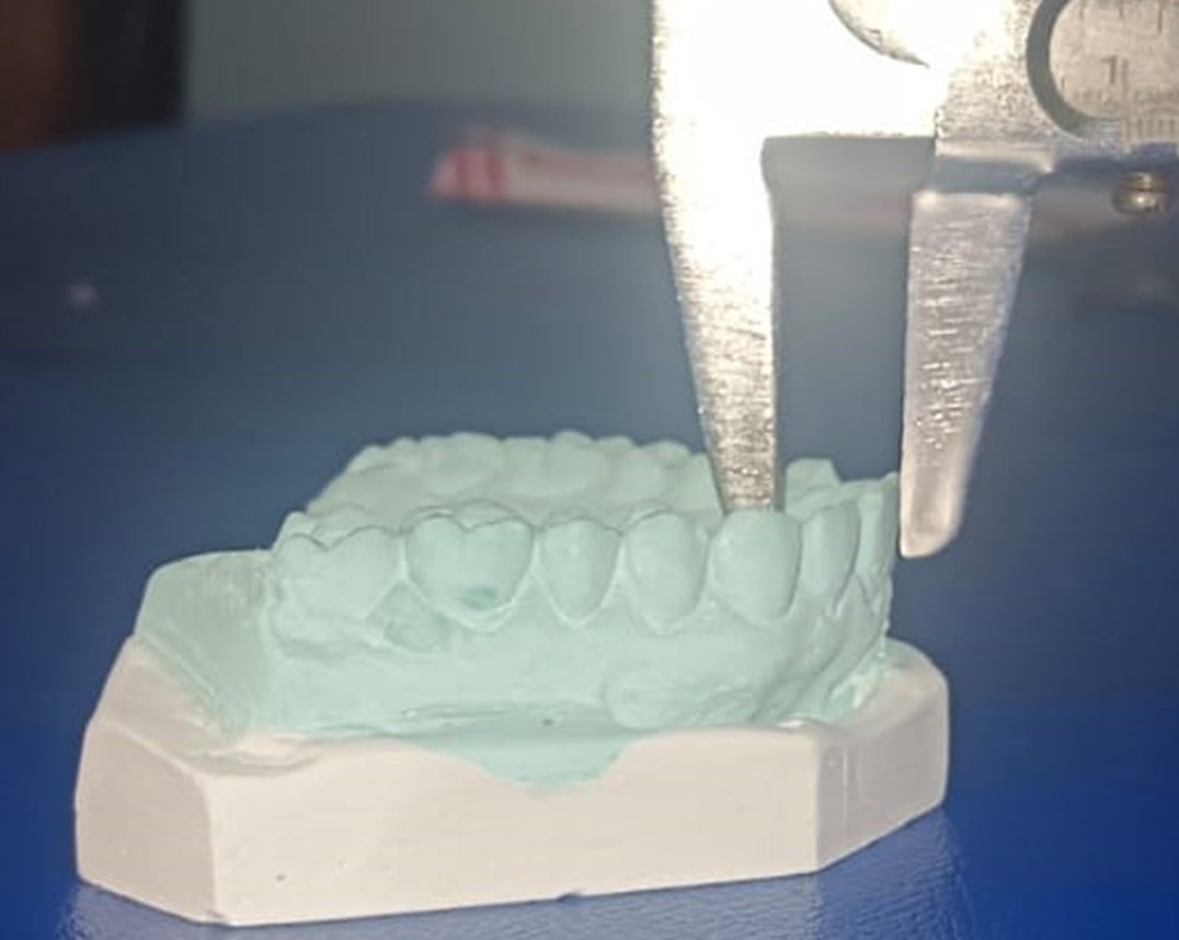
Fig 3: Measuring Horizontal distance using vernier caliper
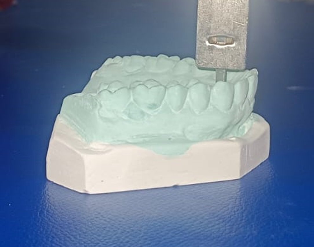
Fig 4: Measuring Vertical distance using vernier caliper
RESULTS
All the statistics have been calculated and computed using IBM Statistical Package for Social Sciences (SPSS) version 20, and the diagrams have been prepared using Microsoft Excel 2019.
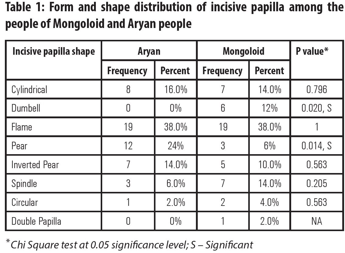
In Mongoloid group, among 50 subjects flame shaped papilla was seen most (n=19) followed by spindle(n=7), cylindrical(n=7), pear(n=6), inverted pear(n=5), dumbbell(n=3), circular(n=2), double papilla is less common(n=1). In Aryan group also, among 50 subjects flame shaped papilla was seen highest (n=19) followed by dumbbell(n=12), cylindrical(n=8), Inverted pear(n=7), spindle(n=3), circular(n=1)(Table 1).
In mongoloid group, Among 50 sample, 30(60%) showing location of the intercanine line in the middle third, 8(16%) in anterior third,12(24%) in the posterior third. In Aryan group, 32(64%) among 50 sample showing location of intercanine line in the middle third of the papilla, 8(16%) in anterior third and10(20%) in the posterior third(Table 2).

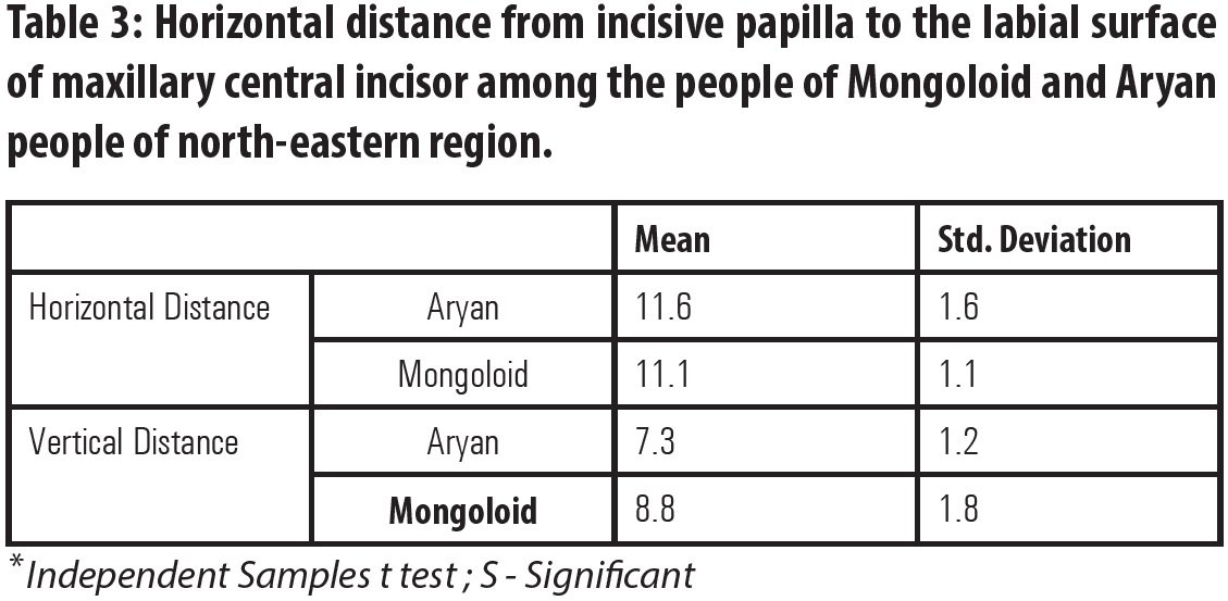
In Mongoloid group, horizontal and vertical distance ranges (11.1±1.1) mm and (8.8±1.8) mm with a mean value of 11.1mm and 8.8mm respectively. However, in Aryan group, horizontal and vertical distance ranges from (11.6±1.6) mm and (7.3±1.2) mm with a mean value of 11.6 mm and 7.3 mm respectively(Table 3).
DISCUSSION
Harper6 in 1949 stated that the position of the incisive papilla remains fairly constant in the edentulous patient since resorption has already taken place in anteroposterior direction.18 So, incisive papilla can use as a guide for placement of anterior teeth in denture. But the shape, location of IP varies in different races. The present study was conducted to measure the form and shape distribution of incisive papilla, location of intercanine line in respect to incisive papilla, horizontal and vertical distance between incisive papilla and central incisor among the North-eastern people of Mongoloid and Aryan origin in order to reduce chairside and lab time. Form and shape of the incisive papilla was determined by marking the papilla in the cast. The location of inter-canine line in respect to anterior, middle, and posterior third of incisive papilla was observed in the photograph by using Adobe photoshop CS3 software. Horizontal Papilla-incisor distance was recorded to the tenth of a millimeter by adjusting the pointed arms of the vernier caliper to contact the reference point at the base of the incisive papilla and the maximum convexity of central incisor. Vertical distance between incisive papilla and central incisor was measured from reference point and the incisal edge of central incisor the point of intersection.
In respect to form and shape of incisive papilla flame shaped papilla was most common in both Mongoloid (38%) and Aryan (38%) group. In Mongoloid group, flame shaped papilla was followed by cylindrical (14%), spindle (14%), pear (12%), inverted pear (10%), dumbell(6%), circular (4%) and least was seen in double papilla (2%). In Aryan group, flame shaped papilla was followed by pear (24%), cylindrical (16%), inverted pear (14%), spindle (6%), circular (2%). Solomon (2012)11 found pear shaped papilla most common in Dravidian population. Kotwal (2024)23 found cylindrical shape of incisive papilla was the most common followed by pear shape in people of jammu region. Ortman (1979)24 found pear shaped papilla, the most common and oval shapes being the next most frequent.
In respect to location of intercanine line, for both Mongoloid and Aryan group of people intercanine line, located mostly on the middle third (60% and 64%) of incisive papilla followed by anterior (24% and 20% respectively) and posterior (16% and 16% respectively) third. Solomon showed inter-canine line passed through the middle of the papilla in 78%, along the base in 14.1 % and in 0.4 % along the anterior border. Tripathi (2019)21 reported that intercanine line passed through the posterior border of the incisive papilla in maximum cases followed by the middle third, middle-posterior, and least through the anterior border of the incisive papilla. Sreshtha et al. (2016)13 reported that intercanine line passed through the middle third of incisive papilla in 52.6% of the subjects, which was closely matched with the study of Lau and Clark (1993)9 whereas, 57.3% subjects had the line passing through the middle third. The canine-papilla-canine line passed through the middle third in 50% of Aryan and 55.3% of Mongoloid group followed by posterior and anterior third. Ehrlich and Gazit (1975)10 found that in 57.6% intercanine lines passed through the center of the papilla. Sawiris (1977)25 stated that in 64% of subjects the intercanine line was situated 1 mm from the center of the papilla. Grove and Christensen (1989)14 found that in 92% of the 50 subjects, the posterior point of the incisive papilla was approximately 3 mm anterior to a line between the distal points of the canines.
In respect to horizontal and vertical distance between incisive papilla and central incisor, the horizontal distance between labial surface of central incisor to reference point of incisive papilla ranges (10-12.2) mm and (10-13.2) mm with a mean value 11.1 mm and 11.6 mm in Mongoloid and Aryan group respectively. Vertical distance between incisal edge of central incisor to reference point of incisive papilla ranges (7-10.6) mm and (6.1-8.5) mm with a mean value 8.8 mm and 7.3 mm in Mongoloid and Aryan group respectively. Ortman and Tsao(1979)24 reported the horizontal distance between most anterior part of the maxillary central incisors and the posterior of the incisive papilla was 12.45 mm. Grave and Becker(1987),26 showed this similar measurement as 12 to 13 mm. Lassila et al.(2001)27 found measurement of the same distance as 12 mm. Sreshtha et al. (2016)13 reported that the distance from the labial surface of maxillary central incisors to the posterior border of the incisive papilla ranged from 9 to 15.9 mm with a mean of 11.59 mm (SD 1.3) in Nepalese population. In contrast, the study done by Isa and Abduladhi28, in Malay, Indian, and Chinese dentate and edentulous subjects, the mean distance from the midpoint of incisive papilla was found to be 9.59 mm. Pachore et al.(2017)2 observed the horizontal distance between maxillary central incisor and midpoint of the papilla was 4-7 mm, 6-8 mm, and 5-8 mm and vertical distance was 4-6 mm in ovoid, square and tapered arch forms. Shah et al. (2014)19 showed that the mean vertical distance of maxillary CI-IP ranged from 6.31 ± 0.74 mm to 7.04 ± 0.87 mm. Maskey et al. (2018)20 reported the mean horizontal distance from the incisal edge of the central incisors to the center of the incisive papilla was 10.04±1.17. Zakiah et al. (2012)29 Grave et al. (1987)26 and Fu et al. (2007)30 reported the similar findings. Hicky and Boucher reported the horizontal distance between incisal edge of the central incisors to the center of the incisive papilla was 8-10mm.16 The distance between the incisal edge of the maxillary central incisors and the posterior of the incisive papilla was 13.58 which was similar the studies done by Soo and Tae (2016)31 and Grave and Becker (1987).26 However, there are a few limitations to the results of this study as well. Firstly, a relatively smaller sample size was included (n=100). Secondly, despite careful calibration, the use of vernier caliper and photographic analysis may be subject to minor observer variations. Impression inaccuracies, cast fabrication errors, and photographic angulation could have influenced the accuracy of landmark identification and measurements.
CONCLUSION
- Flame shaped papilla was the most common in both Mongoloid (38%) and Aryan (38%) group of people.
- For both mongoloid group and Aryan group intercanine line was located in the middle third of the papilla.
- In Mongoloid group horizontal and vertical distance ranges (11.1±1.1) mm and (8.8±1.8) mm respectively. However, in Aryan group, horizontal and vertical distance ranges from (11.6±1.6) mm and (7.3±1.2) mm respectively.
REFERENCES
- Pound E. Recapturing esthetic tooth position in the edentulous patient. J Am Dent Assoc. 1957;55(2):181–191.
- Pachore M et al. An in vivo comparison of vertical and horizontal distance between incisive papilla and incisal edge of maxillary central incisors in dentates with different arch forms. J Clin Diagn Res. 2017 Apr;11(4): ZC97–ZC100.
- Shin YS. Correlation between the size of the incisive papilla and the distance from the incisive papilla to the maxillary anterior teeth. J Dent Sci. 2015; 10:1–5.
- Jayakumar A et al. Prosthodontic significance of incisive papilla: an overview. Acta Sci Dent Sci. 2021 Jun;5(6):1–6.
- Agrawal SK, Singhal R, Pagia S, Kumari K. Evaluation of papillo-incisal distance in different arch forms and with different shapes of incisive papilla in Moradabad population: a descriptive study. J Dent Probl Solut. 2021;8(1):042–046.
- Harper RN. The incisive papilla: the basis of a technique to reproduce the positions of key teeth in prosthodontia. J Prosthet Dent. Year not available.
- Sapkota B, Srii R, Srivastava S, Marla V, Koju S. Assessment of the relationship of incisive papilla to maxillary central incisor and canine–papilla–canine line among the dentate population of Central Nepal. Kathmandu Univ Med J (KUMJ). 2017 Apr–Jun;15(58):150–154.
- Watt DM. Morphological changes in the denture-bearing area following the extraction of maxillary teeth. Br Dent J. 1974; 136:231–235.
- Lau GC, Clark RF. The relationship of incisive papilla to the maxillary central incisors and canine teeth in southern Chinese. J Prosthet Dent. 1993; 70:86–96.
- Erlich J, Gazit E. Relationship of maxillary central incisors and canines to the incisive papilla. J Oral Rehabil. 1975; 2:309–312.
- Solomon EGR, Arunachalam KSJ. The incisive papilla: a significant landmark in prosthodontics. J Indian Prosthodont Soc. 2012;12(4):236–247.
- Schiffman P. Relation of the maxillary canines to the incisive papilla. J Prosthet Dent. 1964; 14:469–472.
- Shrestha S, Joshi SP, Yadav SK. Relationship of incisive papilla to maxillary incisors and canines. J Contemp Dent Pract. 2016;17(4):306–312.
- Grove HR, Christensen LV. Relationship of maxillary canines to the incisive papilla. J Prosthet Dent. 1989; 42:492–496.
- McGee GF. Tooth placement and base contour in denture construction. J Prosthet Dent. 1960; 10:651–657.
- Hickey JC, Boucher CO, Woelfel JB. Responsibility of the dentist in complete dentures. J Prosthet Dent. 1962; 12:637–653.
- Mavroskoufis F, Ritchie GM. Nasal width and incisive papilla as guides for the selection and arrangement of maxillary anterior teeth. J Prosthet Dent. 1981; 45:592–597.
- Guldag MU, Sentut F, Buyukkaplan US. Investigation of vertical distance between incisive papilla and incisal edge of maxillary central incisors. Eur J Dent. 2008 Jul;2(3):161–166.
- Shah M, et al. A comparative study to evaluate the vertical position of maxillary central incisor and canine in relation to incisive papilla line. J Forensic Dent Sci. 2014 May–Aug;6(2):94–99.
- Maskey S, Shrestha R, Yadav R, Yadav NC. Correlation of incisive papilla and the distance from the maxillary anterior teeth. J Med Sci Clin Res. 2018 Oct;6(10):672–674.
- Tripathi S et al. Evaluation of the incisive papilla as a guide to maxillary canine position in the Moradabad population: an anthropometric analysis. Indian J Dent Sci. 2019;11(4):1–5.
- Albuquerque L, Sunila S, De Souza FM. Relationship of maxillary intercanine line to incisive papilla in the population of three South Indian states: a cross-sectional study. World J Dent. 2023 Apr;14(4). doi:10.5005/jp-journals-10015-221.
- Kotwal M, Sharma S, Gupta R, Gautam N, Sharma A. Papillo-incisal distance: a guide to maxillary anterior teeth placement during complete denture fabrication in population of Jammu region. Ann Int Med Dent Res. 2024 Jul–Aug;10(4):94–98.
- Ortman HR, Tsao DH. Relationship of the incisive papilla to the maxillary central incisors. J Prosthet Dent. 1979 Nov;42(5):492–496.
- Sawiris MM. The role of anthropometric measurement in design of complete denture. J Dent. 1977;5(2):141–148.
- sGrave AM, Becker PJ. Evaluation of the incisive papilla as a guide to anterior tooth position. J Prosthet Dent. 1987 Jun;57(6):712–714.
- Lassila LV, Klemetti E, Lassila VP. Position of teeth on the edentulous atrophic maxilla. J Oral Rehabil. 2001; 28:267–272.
- Isa ZM, Abdulhadi LM. Relationship of maxillary incisors in complete dentures to the incisive papilla. J Oral Sci. 2012;54(2):159–163.
- Zakiah M, Isa M, Abdulhadi LM. Relationship of maxillary incisors in complete dentures to the incisive papilla. J Oral Sci. 2012;54(2):159–163.
- Fu PS, Hung CC, Hong JM, Wang JC. Three-dimensional analysis of the occlusal plane related to the hamular–incisive–papilla occlusal plane in young adults. J Oral Rehabil. 2007; 34:136–140.
- Shin SY, Kim TH. Correlation between the size of the incisive papilla and the distance from the incisive papilla to the maxillary anterior teeth. J Dent Sci. 2016;11(2):141–145.
Disclaimer from Guident
The views and opinions expressed in articles published in Guident are solely those of the authors and do not necessarily reflect the views of the editors, the editorial board or the publisher, Ivory India. Guident does not endorse any product, technique or treatment described. Clinical content is intended for qualified dental and medical professionals and is not a substitute for individual clinical judgement. Guident, its editors and the publisher accept no liability for any consequence arising from the use of information published in the journal.
How to cite this article
Dr. Debalina Basak, Dr. Jogeswar Barman. Comparative Evaluation of Horizontal and Vertical Distance Between Maxillary Central Incisors and Incisive Papilla Among the North-Eastern People of Mongoloid and Aryan Origin. Guident. 2026 May;19(6). Available from: https://ivoryindia.in/articles/research/comparative-evaluation-of-horizontal-and-vertical-distance-between-maxillary-central-incisors-and-incisive.html