Evaluation of the Mode of Action of Nonpolymerizable and Polymerizable Desensitizers on Teeth with Scanning Electron Microscopic Analysis
Pandit Deendayal Upadhyay Dental College, Solapur, Maharashtra, India. Dr. Rajendra Avhad (Assistant Professor & HOD), India, Department of Dentistry & Prosthodontics, Crown and Bridge
ABSTRACT
Increased human longevity has led to a rise in dental treatment needs. Majority of partially edentulous patients turning to dental clinics today give more emphasis on fixed prosthesis as compared to removable prosthesis. However, frequently patients exposed to fixed restorative procedures experience discomfort in the prepared teeth either during the treatment or sometimes after cementation of restoration, which they perceive in the form of sensitivity due to which the clinical application of desensitizing agents become prevalent. This study evaluated the nature of action of polymerizable desensitizer, Bis-Block and non-polymerizable glutaraldehyde based GLUMA Desensitizer on the prepared tooth surface by Scanning Electron Microscopic (SEM) Study.
Keywords - Teeth sensitivity ,Polymerizable and Non polymerizable desensitizing agents, Scanning Electron Microscopic (SEM) analysis.
INTRODUCTION
Dentin hypersensitivity is defined as short, sharp pain arising from exposed dentin typically in response to chemical, evaporative, thermal, tactile or osmotic stimuli, which cannot be ascribed to any other form of dental defect or pathology1.
Brannström2 (1964) popularized the ‘hydrodynamic theory’ of dental pain originally proposed by Dr. Kramer in 1955. Brannstrom’s Hydrodynamic theory (1963) speculates that whenever a pain-producing stimulus is applied to the external tooth surface, the fluid flow within the dentinal tubules in either direction stimulates the sensory nerves in dentin or pulp, resulting sensitivity. Alfred Gysi3 (1900) believed that the sensitivity occurred due to fluid movement in the dentinal tubules and suggested for the first time that coagulating the protein content in the outer portion of the tubules can relieve the hypersensitivity.
Richardson, Tao and Pashley (1991)4 reported that on an average approximately 1 to 2 million dentinal tubules are exposed during tooth preparation for a posterior crown. These open dentinal tubules allow fluid flow leading to sensitive dentin. After tooth preparation too, dentin can still become sensitive as a result of interim restoration microleakage and the resultant formation of bacterial byproducts5. Hypersensitivity has been reported even after cementation of definitive prostheses with the most widely used luting agents like zinc phosphate and glass ionomer cement 6,7. This occurs due to the acidic nature of these cements which results in a partial removal of the smear layer. Hence, the cement can enter the dentinal tubules before the luting agent sets8 displacing an equal volume of dentinal fluid in the pulp, which may cause excessive hydrostatic pressure leading to post-cementation sensitivity.
The treatment of hypersensitive teeth should therefore be directed towards reducing the functional diameter of the tubules to limit fluid movement. Several approaches have been proposed to accomplish this objective9, which include
1. Formation of a smear layer by burnishing the exposed tooth surface.
2. Topical application of agents that form insoluble precipitates within
the dentinal tubules.
3. Impregnation of tubules with resins.
4. Application of dentin bonding agents to seal off the tubules.
Application of desensitizing agents is gaining popularity with the advance in dental treatment . This study examined the nature of action of polymerizable desensitizer, Bis-Block and non-polymerizable, glutaraldehyde based GLUMA Desensitizer on the prepared tooth surface by Scanning Electron Microscopic (SEM) Study.
MATERIAL AND METHODS
Attempts were made to standardize the machinery and the procedures throughout the study to minimize the effect of variables on the observations and the result.
1. Selection and preparation of the specimen:
Ninety freshly extracted human molars were selected .These molar teeth were stored in 0.05% sodium azide to prevent microbial growth.The selected molar teeth were cleaned with periodontal scalers under tap water to remove any surface deposits and then stored in distilled water at room temperature, with 0.1% thymol as preservative. Notches were made on the root surface with a diamond point to permit better mechanical locking of teeth in acrylic resin. A permanent mark was made on the tooth root, 1mm below the cementoenamel junction to allow embedding of the specimen upto this level in acrylic resin .
2. Specimen mounting:
Metal molds with internal dimensions of 25mm X 25 mm were used for embedding of teeth specimens. The molds were partially filled with autopolymerizing acrylic resin that was allowed to set . The plane of the surveying table was made parallel to the surveying platform and checked by using spirit level . Small amount of cyanoacrylate was used to position the tooth in its most vertical alignment to the analyzing rod of the surveyor. The mold position was adjusted such that the tooth would be in its center .
The vertical arm was then lowered till the mark on the root surface was in level with the upper rim of the metal mold . The remaining portion of the rings was then filled with autopolymerizing acrylic resin to secure the roots of the extracted teeth (Fig 1). After the resin was polymerized, the analyzing rod was manually separated from the tooth surface. The specimens were stored in distilled water except during tooth preparation.
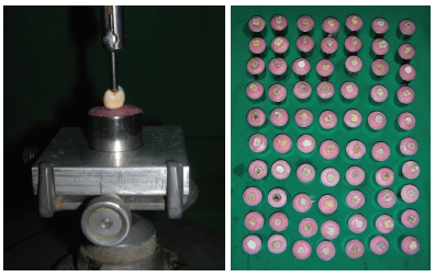
Fig 1: Embedding of tooth in autopolymerizing acrylic resin.
3. Tooth preparation
A wheel shaped diamond point (WR- 13, Dia-Burs, Mani Inc. Japan) was used to prepare the occlusal surface of the tooth. Axial reduction was achieved by using the long round ended tapered diamond instrument (TR-13, Dia - Burs, Mani Inc, Japan). Goodacre10 (2001) recommended a minimum finish line depth of approximately 0.3mm for all metal crowns. Therefore, a 0.5mm chamfer gingival finish line, corresponding to half the diameter of the diamond point at its tip was prepared above the cementoenamel junction. The preparations were finished by using extra fine grit (20-30µ ) round ended tapered diamond abrasive (TR-13EF, Dia-Burs, Mani Inc, Japan). (Fig.2).

Fig 2: Prepared specimens.
The prepared specimens were divided into three groups . Thirty samples were treated for each group.
Group-C –No desensitizing agent applied (control)
Group-B – BisBlock applied
Group-G–GLUMA Desensitizer applied
4. Application of desensitizing agents
The application of desensitizing agents was carried out in following manner:
1. Thirty samples serving as control were left untreated.
2. Thirty samples were treated with GLUMA desensitizer as per manufacturer’s instructions. (fig. 3). A smallest possible amount of GLUMA desensitizer was applied on the prepared tooth surfaces using cotton pellets for 30-60 seconds . Excess liquid was then gently dispersed into a thin layer using a chip syringe. Air was applied until the fluid film had disappeared and the surface was no longer shiny. Then it was rinsed thoroughly with water. Compressed air was not used to avoid desiccations of dentinal surface.
3. Thirty samples were treated with BisBlock desensitizer as per manufacturer’s instructions (fig. 3) .

Fig. 3: Desensitizers used in the study.
- Teeth surface was etched with 32% Phosphoric acid for 15 seconds.
- The etchant was rinsed off and then dried.
- BisBlock was applied for at least 30 seconds.
- Then it was rinsed and the surface was left moist for bonding.
- Then adhesive, ONE-STEP was applied and light cured for 10 seconds using light cure unit CU- 100 RLC on every surface as per manufacturer’s instruction.
5. Processing and study of samples using scanning electron microscope
Teeth were sectioned in mesiodistal direction using a diamond point. From each buccal surface, a sectioned sample of size 5mm length x 5mm width x 3.5mm depth was obtained. Immediately after cleaning of prepared tooth surface, specimens were fixed in 2.5% glutaraldehyde for 12 hrs at 40 C. After fixation, the specimen were washed with phosphate buffer solution and then dehydrated in ascending grades of alcohol (50%, 70%, 90%, and 100%) for 10 minutes each at room temperature. After the specimens were air dried, they were mounted on aluminum stubs with clay. Tooth sample is a non-conductive material. The samples were put into the Gold sputter coating machine (HUMMER VI – A, ANATECH LTD, ALEXANDRIA, VA) in which they (non-conducting samples) were coated with a thin layer of Gold (150-200Aºthick) to make it conductive.(fig. 4). Analysis using a Scanning Electron Microscope (S-3400N Analytical Scanning Electron Microscope) operated at 15 kV was carried out (fig. 5) . Each specimen surface was scanned at desensitizer-dentin interface. Areas showing characteristic features were located and photographed (Fig. 6, 7,8)

Fig. 4: Gold sputter coating machine (HUMMER VI – A, ANATECH LTD, ALEXANDRIA).

Fig. 5: S-3400N Analytical Scanning Electron Microscope
RESULTS
Scanning Electron Microscopic Analysis of the Gluma desensitizer treated dentin revealed that the majority of tubules were obliterated with a thin layer that covered the surface and infiltrated into tubules as plugs (Fig.7 ). Bis-Block forms oxalate crystals deep within the dentinal tubules, (Fig. 8).
DISCUSSION
Tooth hypersensitivity is one of the oldest recorded complaints of discomfort to people. The study of the factors which lead to opening of the dentinal tubules and the treatment methods which can occlude or seal off the exposed dentinal tubules so as to decrease sensitivity , have been explored over the past few decades.
GLUMA Desensitizer: It is non-polymerizable glutaraldehyde resin sealer. According to the manufacturer, It is composed of an aqueous solution containing 5% glutaraldehyde, 35% 2-hydroxyethyl methacrylate (HEMA) and purified water. Glutaraldehyde is a biologic tissue fixative that coagulates and denatures the serum proteins in the dentinal tubule fluid. Hydroxyethyl methacrylate is water soluble that facilitates the deep penetration of glutaraldehyde into the dentinal tubule.
Schupbach P, Lutz F, Finger WJ11 (1997) evaluated the effect of 5% glutaraldehyde and 35% Hydroxyethyl methacrylate on dentin using various microscopic techniques. It was concluded that glutaraldehyde can intrinsically block dentinal tubules thereby reducing sensitivity.


Fig 6: Scanning Electron Microscopy of control group.

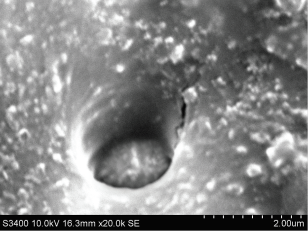
Fig.7: Scanning Electron Microscopy of BisBlock group.

Fig. 8: Scanning Electron Microscopy of Gluma desensitizer group.
Arris CAG et al12 (2004) evaluated the features of dentinal tubules occlusion following application of three commercially available desensitizing agents: Oxa-Gel, Gluma Desensitizer and Nupro Gel. They stated that according to Scanning electron microscopic analysis all desensitizing agents were able to occlude the dentinal tubules. In that clinical study the primer of the original Gluma Adhesive system (currently marketed as Gluma Desensitizer), the desensitizing solution was applied to crown preparations. They concluded that Gluma primer reduce sensitivity through a protein denaturation process with concomitant changes in dentin permeability. This theory recently has been supported by studies using conofocal microscopy13, which found formation of transversal septa occluding the dentinal tubules following application of Gluma desensitizer. SEM analysis in this study also found that Gluma Desensitizer infiltrates into the tubules as plugs and form thin layer over the dentin occluding the entrance of the tubules confirming the results of above mentioned studies.
Post-operative sensitivity is frequently encountered with the use of adhesives that require the acid etching of vital dentin14. The application of these total-etch adhesives can be technique-sensitive, since wet bonding is required for full expansion and optimal hybridization of the demineralized collagen matrix. The difficulty in bonding is further complicated by the intrinsic wetness of vital deep dentin after removal of the smear layer and the increased permeability associated with the simplified version of these adhesives. Incomplete sealing and continuous transudation of dentinal fluid through open dentinal tubules before polymerization of the adhesive may result in entrapment of water-filled blisters along the adhesive interface. Compression of these blisters during mastication may cause, within the dentinal tubules, rapid fluid movement that activates the intradental A8 nerve fibers, which results in post-operative sensitivity. One way of relieving post-operative sensitivity clinically is the adjunctive use of oxalate desensitizers on acid-etched dentin prior to adhesive application (Pashley et al., 200115; Tay et al., 2003b16).
Bis-Block Desensitizer
According to the manu facturer, BisBlock is an oxalate De sensitizer, containing < 5% Oxalic Acid. It reacts with the Calcium of dentine and forms calcium oxalate crystals, which occlude the dentinal tubules and thus reduce hyper sensitivity,17,18Depletion of calcium ions from the surface dentin forces the oxalate ions to diffuse further down into the dentinal tubule, until calcium ions are encountered for reaction. The calcium oxalate crystals that are formed result in subsurface tubular occlusion and reduction in the hydraulic conductance of dentin. SEM analysis in this study also shows calcium oxalate crystals in the opening of the dentinal tubules and small aggregates on the surfaces, confirming the results of above mentioned studies.
However, the solubility of calcium oxalate is affected by pH, since the anion is the conjugate base of a weak acid (Kotz and Treichel, 1999). Preliminary screening of the compatibility of oxalate desensitizers with total-etch adhesive systems revealed complete compatibility with One-Step (Bisco) and Single Bond (3M ESPE)14. In this study, One-step is used. One-step also mechanically occludes open tubules with resin, thus sealing them from oral environment.
Scanning electron microscopic observation of untreated prepared tooth surface, i.e. control group showed no changes on the dentinal surface.
CONCLUSION
The scanning electron microscopy revealed that there is definite interaction of the Gluma desensitizer with exposed dentinal tubules, as it infiltrates into the tubules as plugs and forms thin layer over the dentin occluding the entrance of the tubules.The scanning electron microscopy also revealed the definite interaction of the Bis-Block with exposed dentinal tubules. There is formation of calcium oxalate crystals deep inside the dentinal tubules, an adhesive layer over the dentin and hybrid layer interposed in between them.
Clinicians deal with crown and bridge restorative procedures on a regular basis. Desensitizing agents should be used routinely on all vital abutment teeth to be restored with fixed restorations not only to prevent dentin hypersensitivity but equally important to minimize bacterial contamination to reach the pulp.
REFERENCES
- Addy M. : Etiology and clinical implications of dentine hypersensitivity. Dent Clin North Am 1990; 34(3): 503-14.
- Brannström M, Aström A. A study on the mechanism of pain elicited from the dentin. J Dent Res 1964; 43:619-26.
- Gysi A. An attempt to explain the sensitiveness of dentin. Br J Dent Sci 1900; 43:865-8.
- Richardson D, Tao L, Pashley DH. Dentin permeability; effect of crown preparation. Int J Prosthodont 1991; 4:219-25.
- Bergenholtz G. Inflammatory response of the dental pulp to bacterial irritation. J Endod 1981;7:100-4.
- Kern M, Kleimeier B, Schaller H, Strub JR. Clinical Comparison of postoperative sensitivity for a glass ionomer and a zinc phosphate luting cement. J Prosthet Dent 1996; 75:159-62.
- Bebermeyer RD, Berg JH. Comparison of patient-perceived post cementation sensitivity with glass ionomer and zinc phosphate cements. Quintessence Int 1994; 25:209-14.
- Zaimoglu A, Kevser A. An evaluation of smear layer with various desensitizing agents after tooth preparation. J Prosthet Dent 1992; 68:450-457.
- Trowbridge HO, Silver DR. A review of current approaches to In-office management of tooth hypersensitivity. Dent Clin North Am 1990; 34(3):561-81.
- Goodacre CJ, Campagni WV, Aquilino SA. Tooth preparations for complete crowns: An art form based on scientific principles. J Prosthet Dent 2001; 85:363-76.
- Schupbach P, Lutz F, Finger WJ : Closing of dentinal tubules by Gluma desensitizer, Eur J Oral Sci. 1997;105: 414-21.
- Arrais CAG, Chan DCN Giannini M. Effects of desensitizing agents on dentinal tubule occlusion. J Appl Oral Sci 2004; 12(2):144-8.
- Schupbach P, Lutz F, Finger WJ. Closing of dentinal tubules by Gluma desensitizer. Eur J Oral Sci 1997; 105:414-21.
- C.K.Y. Yiu, N.M. King, B.I. Suh, L.J. Sharp, R.M. Carvalho, D.H. Pashley, and F.R. Tay: Incompatibilities of oxalate Desensitizers with Acidic, Fluoride- containing Total etch Adhesives. J Dent Res 84: 730-735, 2005.
- Pashley DH, Carvalho RM, Pereira JC, Villanueva R, Tay FR: The use of oxalate to reduce dentin permeability under adhesive restorations. Am J dent 2001; 14:89- 94.
- Tay FR, Pashley DH, Mak YF, Carvalho RM, Lai SC, Suh BI (2003b): integrating oxalate desensitizers with total etch two steps adhesive. J Dent Res 82: 703-707.
- Richardson DW, Tao L, Pashley DH. Bond strengths of luting cements to potassium oxalate- treated dentin. J Prosthet Dent 1990; 63: 418-22.
- Pashley DH: Dentin permeability, dentin sensitivity, and treatment through tubules occlusion. J Endod 1986; 12: 465-474.
Disclaimer from Guident
The views and opinions expressed in articles published in Guident are solely those of the authors and do not necessarily reflect the views of the editors, the editorial board or the publisher, Ivory India. Guident does not endorse any product, technique or treatment described. Clinical content is intended for qualified dental and medical professionals and is not a substitute for individual clinical judgement. Guident, its editors and the publisher accept no liability for any consequence arising from the use of information published in the journal.
How to cite this article
Dr. Ranjukta Sar Avhad, Dr. V M Government medical college and Chatrapati Shivaji Maharaj Sarovpchar Rugnalya Solapur. Maharashtra. Evaluation of the Mode of Action of Nonpolymerizable and Polymerizable Desensitizers on Teeth with Scanning Electron Microscopic Analysis. Guident. 2019 Mar;12(4). Available from: https://ivoryindia.in/articles/research/evaluation-of-the-mode-of-action-of-nonpolymerizable-and-polymerizable-desensitizers-on-teeth-with-scanning.html