Tilted Implantswiss Implant Placement in Posterior Maxilla with Customized Prosthesis: A Case Report
Wisdom Dental Clinics ®, Jaipur, India
ABSTRACT
The success of dental implant procedures is dependent on many factors. Overall medical status, health, and economic considerations can be limiting factors in determining which of our patients are appropriate candidates. Additionally, the time required for some implant treatments, and the need to perform bone grafting and augmentative procedures in a staged manner often deter patients from proceeding with implant-borne prosthetic rehabilitation.
Recently, numerous alternatives have been introduced and developed to circumvent anatomic limitations and the need for grafting or nerve repositioning procedures and their attendant economic costs, treatment times and morbidities. The use of shorter implants, ridge splitting/ridge expansion/ and densification surgeries using specially designed burs or piezotomes, and angled prosthetic components allowing for angulated or tilted implant placement, in deficient areas are challenging traditional dogma.
The purpose of this article was to report a case utilizing tilted angulated implants in the posterior maxilla thereby facilitating fabrication of a multi-unit implant retained fixed dental prosthesis.
Keywords: Angulated Abutments, Angulated Implants, Co-axis, Labially Inclined
INTRODUCTION
Placement and restoration of multiple implants in the maxilla create a host of challenges unique to its anatomic and aesthetic requirements.1,2 Despite bone grafting, computer aided implant positioning software and surgical guides, the labial inclination of the anterior maxilla makes vertical placement of implants difficult and forces the practitioner to labially incline/tilt the implants.3,4,5
Traditional implants are intentionally tilted to achieve longer length, to place the implant in maximal bone, and to avoid vital anatomic structures such as maxillary sinuses, inferior alveolar canal, nasal floor, submandibular/sublingual spaces or mental foramen.6
Pterygomaxillary implants have been used for more than 25 years and were one of the first tilted implant techniques.7 The study of Balshi et al.,8,9 and the study of Valero´n and Valero´n 10support the use of pterygomaxillary implants in the resorbed maxilla. Zygomatic implants have been used for 20 years, are also tilted to achieve better bone anchorage and longer implant length.7 Many studies have concluded that they are predictable and a viable treatment option for severe maxillary resorption.11,12, 13
Another category is the distally tilted posterior implants used for the “All on four” approach.14 They aid in increasing the antero-posterior spread of implants, decreasing the length of the cantilever, increase the posterior extension of the prostheses and decrease the stresses on the implants.7,14 Studies by Krekmanov et al.,15 Menini et al.,16 Aparcio et al.,17 have concluded that tilted implants are successful and are not associated with increased bone loss compared to axially/ vertically placed implants.
Tilted implants are a successful alternative to bone augmentation and nerve repositioning procedures and aid in decreasing the treatment time, expenses and morbidity associated with complicated surgical procedures.6
Tilted implants may make it necessary to use angled abutments to achieve a parallel path of insertion/ removal of the definitive prosthesis.6,7 These abutments improve the trajectory of the emergence profile as well as allow a degree of parallelism with implants placed in the posterior maxilla.5,7,8,9 Parallelism of anterior and posterior implants increases aesthetically favorable treatment options while optimizing the biomechanics of the definitive restoration and simplifying fabrication techniques.4,5 Eger et al.,18 Sethi et al.,19,20 have concluded in their studies that good function and aesthetics can be achieved by restoring tilted implants with angled abutments.
Recently, a novel implant design, which permits correction of the restorative trajectory of the tilted implant without the use of angled abutments, has been introduced. The angulation of the implant is at the neck below the crestal bone permitting interference free fabrication of the prosthesis. The angle correction feature of the implant improves parallelism of multiple implants and the path of insertion of the future restoration. These implants also facilitate the use of standard implant abutments.
The purpose of this article was to report a case utilizing tilted angulated implants in the posterior maxilla thereby facilitating fabrication of a multi-unit implant retained fixed dental prosthesis.
CASE REPORT
A 75-year-old male present to clinic with the chief complaint of poor aesthetics, missing maxillary posterior teeth 13,14,15,16,17. Medical history was taken but nothing was abnormally detected. On intraoral examination patient had missing right maxillary teeth. The reason for tooth loss is extraction due to presence of dentigerous cyst, after extraction residual defect developed. [Figure 1]
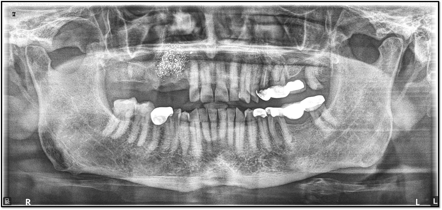
Figure 1: Iodoform dressing after tooth extraction
After 3 months patient was willing for fixed prosthesis to replace missing upper tooth to fulfil both masticatory function and aesthetics. [Figure 2]

Figure 2: CBCT of right maxillary
Clinical and Radiographic evaluation showed adequate labial bone. Routine blood investigations were done. It was planned to restore with implant retained prosthesis. ImplantSwiss Bone level implants were selected based on CBCT evaluation. Following an injection of 2% lidocaine with 1: 80,000 anaesthetic agent in the area of the missing teeth, free hand implant placement was planned. Mid crestal incision was given with incision and full thickness flap was raised and underlying bone was inspected. An osteotomy was performed using lance drill in the edentulous span. A 2.0mm diameter drill was used to make the initial osteotomy till a depth of 10 mm. Subsequent drilling was done till diameter of 3.2 mm and implant insertion done.3 ImplantSwiss implant of size 3.7 X 14 mm,4.3 X14, 4.3X8 were placed. In the osteotomy site with torque ratchet till the insertion torque of 40Ncm and ISQ of more than 75 was achieved. Suturing was done using 3-0 silk suture. [Figure 3]

Figure 3: OPG after implant placement
Antibiotic and anti-inflammatory drugs were prescribed and post operative instructions were given. Patient was recalled after one week for suture removal
Post-operative after 2 months ISQ test done again, which showed excellent biological stability with reading measuring in between 84-87. Thereby showing excellent secondary stability or osseointegration. Post-operative OPG was suggested after 2 months to check hard tissue architecture around ImplantSwiss implants [Figure 4]. Excellent soft tissue healing was seen around ImplantSwiss implant.

Figure 4: OPG after 2 months
Digital impression was made by CEREC Workflow (Primescan). Implant scan bodies were used & it was planned to give screw retained crown by using Implantswiss implant Ti-Base. Sintering of Meso Sirona done using SpeedFire. Now Implantswiss implant Ti -Base Swiss was cemented extraorally with resin-based cement to prepared crown. OPG showed Implant & Prosthesis in perfect 3-D position [Figure 5] ensuring very good prognosis. Occlusion & proximal contacts were verified.

Figure 5: OPG after prosthesis placement
Occlusion was checked and patient was satisfied and happy with this prosthesis [Figure 6 a, b, c].


Figure 6: Post-operative image
DISCUSSION
Tilting of implants facilitates the placement of longer implants, reduction of the cantilever length, achievement of better bone anchorage and better antero-posterior spread of implants.6,7 Tilted implants, which support the prostheses, also aid in stress distribution.24 Many finite element analysis,25-29 animal studies,30,31,32 and clinical studies14,15 have concluded that there is no significant difference in success and survival rates, bone loss and bone stresses between axially placed and tilted implants.
Accurate 3-dimensional implant placement and achievement of implant parallelism is challenging in the maxilla due to its morphology and anatomy.1-6 Tilted implants are unavoidable in the anterior maxilla due to osseous labial proclination.1-6 This often results in screw access that is directed on the facial surface of the restorations, compromising the aesthetics and strength of the definitive restoration. Tilted implants require the use of angled abutments to redirect the restorative platform of the implant lingually to achieve the desired aesthetics.6-7 Adjustments to the angled abutment may be required to reduce bulk and achieve the desired aesthetics.1,6 Adjusting the angled abutment may weaken the abutment and eventually lead to fracture of the definitive restoration.6
The use of the novel angulated implants platform in this case, aided in achieving multi-implant parallelism without bone grafting and eliminated the need to use angled abutments. These implants facilitate the use of standard implant abutments for fabrication of a tough and fracture resistant cement retained fixed dental prostheses. The use of these implants aided in achieving an ideal implant restorative platform position. They allowed additional implant supported abutments to splint, support and retain the prosthesis, the reduction of additional implant componentry for impressions and prosthesis fabrication, decrease in prosthesis and surgical costs, and increased patient acceptance.
Studies on tapered, roughened-surfaced implants with a novel angled prosthodontic platform, conducted by Brown and Payne22,23 and Vandeweghe et al.,5 report the successful use of these novel implants for immediate replacement and immediate restoration of single teeth in the esthetic zone, but they are short-term studies with only 1 year of follow up time.
Additional randomized controlled studies and more long-term clinical trials are necessary to evaluate the durability of these implants and restorations fabricated on multiple placements.
Fixed dental implant supported prostheses can be designed to be cement retained or screw retained. Screw retention was introduced in early 1980’s to facilitate retrievability of failing implant restorations.33 Screw loosening has been reported as a major problem with screw-retained restorations.33-39 Sectioning and soldering procedures are routinely required for screw-retained prostheses.33 In addition, screw retained restorations have some substantial disadvantages compared to cement retained restorations including increased costs, greater complexity of components and laboratory procedures, increased chair-side time and compromised aesthetics.33,34 Screw retained prostheses are usually indicated when there is decreased vertical restorative space and/or when retrievability is desired.33
The purpose of this article was to report a case utilizing tilted angulated implants in the posterior maxilla thereby facilitating fabrication of a multi-unit implant retained fixed dental prosthesis.
CONCLUSION
The novel angulated implants may be a viable alternative to traditional implants used along with angled abutments for replacing and restoring the missing teeth in both the maxilla and the mandible. Further studies are needed to evaluate the success and survival rates of the newly designed implants.
REFERENCES
- Kurtzman GM, Dompkowski DF, Mahler BA, Howes DG. Off-axis implant placement for anatomical considerations using the Co-axis implant. Inside Dentistry. 2008; 5: 96–102. [Google Scholar]
- Schneider AL, Kurtzman GM. Restoration of divergent free-standing implants in the maxilla. J Oral Implantol. 2002; 28: 113–116. [PubMed] [Google Scholar]
- Cavallaro J Jr, Greenstein G. Angled implant abutments: a practical application of available knowledge. J Am Dent Assoc. 2011; 142: 150–158. [PubMed] [Google Scholar]
- De Avila ED, de Molon RS, de Barros-Filho LA, de Andrade MF, Mollo Fde A Jr, de Barros LA. Correction of Malpositioned Implants through PeriodontalSurgery and Prosthetic Rehabilitation Using Angled Abutment. Case Rep Dent. 2014; 2014: 702630. [PMC free article] [PubMed] [Google Scholar]
- Vandeweghe S, Cosyn J, Thevissen E, Van den Berghe L, De Bruyn H. A 1-year prospective study on Co-Axis implants immediately loaded with a full ceramic crown. Clin Implant Dent Relat Res. 2012; 14 Suppl 1: e126–e138. [PubMed] [Google Scholar]
- Tahmasebi S, Nicolopoulos Costa. Modern implants from a different angle. Dental tribune Middle east and Africa edition 2015; Jan-Feb:24-25. Available at: [http://www.dental-tribune.com/articles/specialities/implantology/21668_modern_implants_from_a_different_angle.html.]
- Graves S, Mahler BA, Javid B, Armellini D, Jensen OT. Maxillary all-on-four therapy using angled implants: a 16-month clinical study of 1110 implants in 276 jaws. Dent Clin North Am. 2011; 55: 779–794. [PubMed] [Google Scholar]
- Balshi TJ, Wolfinger GJ, Balshi SF 2nd. Analysis of 356 pterygomaxillary implants in edentulous arches for fixed prosthesis anchorage. Int J Oral Maxillofac Implants. 1999; 14: 398–406. [PubMed] [Google Scholar]
- Balshi SF, Wolfinger GJ, Balshi TJ. Analysis of 164 titanium oxide-surface implants in completely edentulous arches for fixed prosthesis anchorage using the pterygomaxillary region. Int J Oral Maxillofac Implants. 2005; 20: 946–952. [PubMed] [Google Scholar]
- Valerón JF, Valerón PF. Long-term results in placement of screw-type implants in the pterygomaxillary-pyramidal region. Int J Oral Maxillofac Implants. 2007; 22: 195–200. [PubMed] [Google Scholar]
- Ahlgren F, Størksen K, Tornes K. A study of 25 zygomatic dental implants with 11 to 49 months' follow-up after loading. Int J Oral Maxillofac Implants. 2006; 21: 421–425. [PubMed] [Google Scholar]
- Aparicio C, Ouazzani W, Garcia R, Arevalo X, Muela R, Fortes V. A prospective clinical study on titanium implants in the zygomatic arch for prosthetic rehabilitation of the atrophic edentulous maxilla with a follow-up of 6 months to 5 years. Clin Implant Dent Relat Res. 2006; 8: 114–122. [PubMed] [Google Scholar]
- Bedrossian E, Rangert B, Stumpel L, Indresano T. Immediate function with the zygomatic implant: a graftlesssolution for the patient with mild to advanced atrophy of the maxilla. Int J Oral Maxillofac Implants. 2006; 21: 937–942. [PubMed] [Google Scholar]
- Maló P, Nobre Mde A, Petersson U, Wigren S. A pilot study of complete edentulous rehabilitation with immediate function using a new implant design: case series. Clin Implant Dent Relat Res. 2006; 8: 223–232. [PubMed] [Google Scholar]
- Krekmanov L, Kahn M, Rangert B, Lindström H. Tilting of posterior mandibular and maxillary implants for improved prosthesis support. Int J Oral Maxillofac Implants. 2000; 15: 405–414. [PubMed] [Google Scholar]
- Menini M, Signori A, Tealdo T, Bevilacqua M, Pera F, Ravera G, et al. Tilted implants in the immediate loading rehabilitation of the maxilla: a systematic review. J Dent Res. 2012; 91: 821–827. [PubMed] [Google Scholar]
- Aparicio C, Perales P, Rangert B. Tilted implants as an alternative to maxillary sinus grafting: a clinical, radiologic, and periotest study. Clin Implant Dent Relat Res. 2001; 3: 39–49. [PubMed] [Google Scholar]
- Eger DE, Gunsolley JC, Feldman S. Comparison of angled and standard abutments and their effect on clinical outcomes: a preliminary report. Int J Oral Maxillofac Implants. 2000; 15: 819–823. [PubMed] [Google Scholar]
- Sethi A, Kaus T, Sochor P. The use of angulated abutments in implant dentistry: five-yearclinical results of an ongoing prospective study. Int J Oral Maxillofac Implants. 2000; 15: 801–810. [PubMed] [Google Scholar]
- Sethi A, Kaus T, Sochor P, Axmann-Krcmar D, Chanavaz M. Evolution of the concept of angulated abutments in implantdentistry: 14-year clinical data. Implant Dent. 2002; 11: 41–51. [PubMed] [Google Scholar]
- Sorrentino R, Gherlone EF, Calesini G, Zarone F. Effect of implant angulation, connection length, and impressionmaterial on the dimensional accuracy of implant impressions: an in vitro comparative study. Clin Implant Dent Relat Res. 2010; 12 Suppl 1: e63–e76. [PubMed] [Google Scholar]
- Vojdani M, Torabi K, Ansarifard E. Accuracy of different impression materials in parallel and nonparallel implants. Dent Res J (Isfahan) 2015; 12: 315–322. [PMC free article] [PubMed] [Google Scholar]
- Brown SD, Payne AG. Immediately restored single implants in the aesthetic zone of the maxilla using a novel design: 1-year report. Clin Oral Implants Res. 2011; 22: 445–454. [PubMed] [Google Scholar]
- Zampelis A, Rangert B, Heijl L. Tilting of splinted implants for improved prosthodontic support: a two-dimensional finite element analysis. J Prosthet Dent. 2007; 97 (6 Suppl): S35–S43. [PubMed] [Google Scholar]
- Cruz M, Wassall T, Toledo EM, da Silva Barra LP, Cruz S. Finite element stress analysis of dental prostheses supported by straight and angled implants. Int J Oral Maxillofac Implants. 2009; 24: 391–403. [PubMed] [Google Scholar]
- Bellini CM, Romeo D, Galbusera F, Agliardi E, Pietrabissa R, Zampelis A, et al. A finite element analysis of tilted versus nontilted implantconfigurations in the edentulous maxilla. Int J Prosthodont. 2009; 22: 155–157. [PubMed] [Google Scholar]
- Las Casas EB, Ferreira PC, Cimini CA Jr, Toledo EM, Barra LP, Cruz M. Comparative 3D finite element stress analysis of straight and angled wedge-shaped implant designs. Int J Oral Maxillofac Implants. 2008; 23: 215–225. [PubMed] [Google Scholar]
- Markarian RA, Ueda C, Sendyk CL, Laganá DC, Souza RM. Stress distribution after installation of fixed frameworks with marginal gaps over angled and parallel implants: a photoelasticanalysis. J Prosthodont. 2007; 16: 117–122. [PubMed] [Google Scholar]
- Bellini CM, Romeo D, Galbusera F, Taschieri S, Raimondi MT, Zampelis A, et al. Comparison of tilted versus nontilted implant-supportedprosthetic designs for the restoration of the edentuous mandible: a biomechanical study. Int J Oral Maxillofac Implants. 2009; 24: 511–517. [PubMed] [Google Scholar]
- Celletti R, Pameijer CH, Bracchetti G, Donath K, Persichetti G, Visani I. Histologic evaluation of osseointegrated implants restored in nonaxial functional occlusion with preangled abutments. Int J Periodontics Restorative Dent. 1995; 15: 562–573. [PubMed] [Google Scholar]
- Barbier L, Schepers E. Adaptive bone remodeling around oral implants under axial and nonaxial loading conditions in the dog mandible. Int J Oral Maxillofac Implants. 1997; 12: 215–223. [PubMed] [Google Scholar]
- Francetti L, Romeo D, Corbella S, Taschieri S, Del Fabbro M. Bone level changes around axial and tilted implants in full-arch fixed immediate restorations. Interim results of a prospective study. Clin Implant Dent Relat Res 2012; 14: 646–654. [PubMed] [Google Scholar]
- Hebel KS, Gajjar RC. Cement-retained versus screw-retained implant restorations: achieving optimal occlusion and esthetics in implant dentistry. J Prosthet Dent. 1997; 77: 28–35. [PubMed] [Google Scholar]
- Misch CE. Contemporary implant dentistry. 3th ed. St Louis: Mosby, Year Book Inc; 1993. pp. 651–685. [Google Scholar]
- Kallus T, Bessing C. Loose gold screws frequently occur in full-arch fixed prostheses supported by osseointegrated implants after 5 years. Int J Oral Maxillofac Implants. 1994; 9: 169–178. [PubMed] [Google Scholar]
- Jemt T, Laney WR, Harris D, Henry PJ, Krogh PH Jr, Polizzi G, et al. Osseointegrated implants for single tooth replacement: a 1-year report from a multicenter prospective study. Int J Oral Maxillofac Implants. 1991; 6: 29–36. [PubMed] [Google Scholar]
- Laney WR, Jemt T, Harris D, Henry PJ, Krogh PH, Polizzi G, et al. Osseointegrated implants for single-tooth replacement: progressreport from a multicenter prospective study after 3 years. Int J Oral Maxillofac Implants. 1994; 9: 49–54. [PubMed] [Google Scholar]
- Jemt T, Pettersson P. A 3-year follow-up study on single implant treatment. J Dent. 1993; 21: 203–208. [PubMed] [Google Scholar]
- Carlson B, Carlsson GE. Prosthodontic complications in osseointegrated dental implanttreatment. Int J Oral Maxillofac Implants. 1994; 9: 90–94. [PubMed] [Google Scholar]
Disclaimer from Guident
The views and opinions expressed in articles published in Guident are solely those of the authors and do not necessarily reflect the views of the editors, the editorial board or the publisher, Ivory India. Guident does not endorse any product, technique or treatment described. Clinical content is intended for qualified dental and medical professionals and is not a substitute for individual clinical judgement. Guident, its editors and the publisher accept no liability for any consequence arising from the use of information published in the journal.
How to cite this article
Dr. Gaurav Gupta, Dr. D.K. Gupta, Dr. Devanshi Sankhla. Tilted Implantswiss Implant Placement in Posterior Maxilla with Customized Prosthesis: A Case Report. Guident. 2023 Sep;16(10). Available from: https://ivoryindia.in/articles/implantology/tilted-implantswiss-implant-placement-in-posterior-maxilla-with-customized-prosthesis-a-case-report.html