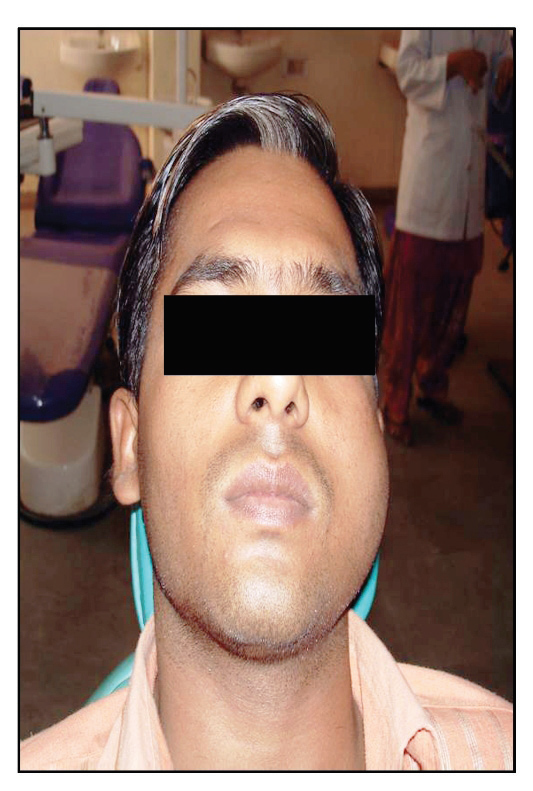
Odontogenic Keratocyst - Cyst / Tumor?
K.D Dental College and Hospital MAthura, U.P, India
INTRODUCTION
Odontogenic Keratocyst (OKC) has been defined as a benign uni or multicystic intraosseous cyst of odontogenic origin, with a characteristic lining of parakeratinized stratified squamous epithelium and potentially aggressive, infiltrative behavior. It may be solitary or multiple. It exhibits paradoxical behavior and may have clinicopathological features of both simple cysts and benign neoplasms.
Odontogenic keratocysts (OKCs) are developmental odontogenic cysts of epithelial origin, first identified and described in 1876 and further characterized by Phillipsen in 1956. Pindborg and Hansen suggested the histologic criteria necessary to diagnose OKC in 1962.
In 2005, the WHO designated OKC as KCOT based on its behavior (locally destructive, high recurrence rate), histopathology (formation of daughter cyst and mitotic figures in the suprabasal layers of the lesional epithelium) and genetics (loss of function of tumor suppressor gene PATCH).
Most commonly occurring bilateral cyst/ lesion is OKC and is more likely to be associated with Nevoid basal cell carcinoma syndrome (NBCCS) than its solitary counterpart. Bilateral (crossing the midline) occurrence of other jaw cysts is extremely rare.
The Journey of classifications and nomenclature of OKC is as follows.1
- Dental cyst (John Hunter 1774),
- Dermoid Cyst ( Mikulicz 1876)
- Primordial cyst (Robinson 1945)
- Keratocystoma (Shear)
- Odontogenic keratocyst ( Philisen 1956 & Pindborg and Hansen 1963)
- Benign neoplasm (Toller 1967)
- Odontogenic keratocyst (WHO 1971)
- True benign cystic epithelial neoplasm (Ahlfors 1984)
- Odontogenic keratocyst (WHO 1992)
- keratocystic odontogenic tumor (Benign neoplasm) (WHO 2005)
- Odontogenic keratocyst (WHO 2017)
CLINICAL FEATURES
KCOTs comprise approximately 11% of all cysts of the jaws6. They occur most commonly in the mandible, especially in the posterior body and ramus regions7. They almost always occur within bone, although a small number of cases of peripheral KCOT have been reported. Patients may present with swelling, pain and discharge or may be asymptomatic. Distinctive clinical features include a potential for local destruction and a tendency for multiplicity, especially when the lesion is associated with nevoid basal cell carcinoma syndrome (NBCCS) or Gorlin-Goltz syndrome. KCOTs have a high recurrence rate, reportedly between 25% and 60% (when associated with NBCCS, the recurrence rate is about 82%)8. In addition to multiple KCOTs, NBCCS is also characterized by nevoid basal cell carcinomas, bifid ribs, calcification of the falx cerebri, frontal bossing, multiple epidermoid cysts and medulloblastoma.
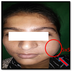
Extraoral view showing swelling on the left side of the face
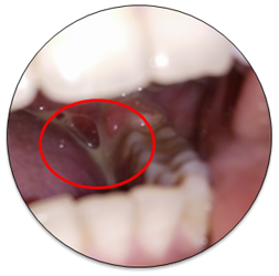
Intraoral view of left side showing draining abscess.
KCOT: THE NEOPLASM
In 1967, Toller suggested that the OKC may be regarded as a benign neoplasm rather than a conventional cyst based on its clinical behaviour. In 1984, Ahlfors and others suggested that “if the OKC were recognized as a true, benign cystic epithelial neoplasia, the question of modified treatment schedules would be raised.”
• Behaviour: As described earlier, the KCOT is locally destructive and highly recurrent.
• Histopathology: Studies such as that by Ahlfors and others show the basal layer of the KCOT budding into connective tissue. In addition, WHO notes that mitotic figures are frequently found in the suprabasal layers.
• Genetics: PTCH (“patched”), a tumour suppressor gene involved in both NBCCS and sporadic KCOTs, occurs on chromosome 9q22.3-q31.
RADIOGRAPHIC FEATURES
Radiographically KCOTs may appear as small, round or ovoid unilocular radiolucencies or may be larger with scalloped margins. The radiolucency tends to be well demarcated with distinct sclerotic margins, but may be diffuse in parts. There is displacement of impacted or erupted teeth, root resorption (10%-15%), root displacement or extrusion of the erupted teeth.
CT scanning may be helpful in detecting cortical perforation and assessment of soft tissue involvement especially in patients with NBCCS–related KCOTs. Contrast enhanced MRI may provide more detailed information.
RADIOLOGICAL TYPES OF KERATOCYST
1. Replacement type : Cyst which forms in the place of normal teeth.
2. Envelopmental type: Cyst which embraces an adjacent unerupted tooth.
3. Extraneous type: Cyst which occur in ascending ramus away from the teeth.
4. Collateral type : Cyst which occurs adjacent to the root of teeth which are indistinguishable radiologically from lateral periodontal cyst.

Panoramic radiograph showing aggressive behavior of OKC with multiple cystic lesions in Non- syndromic patient.

DIFFERENTIAL DIAGNOSIS
- Clinically : amleoblastoma , Dentigerous cyst
- Histologically : myxoma, ameloblastoma, central giant cell granuloma, odontogenic cysts.
- Radiographically : Dentigerous cyst (40%), Residual cysts, radicular cyst, Lateral periodontal cysts (25%), Primordial cyst (25%), Globulomaxillary cyst (10%), Unicystic ameloblastoma, A-V malformation, Fibroosseous lesion at initial stages.
HISTOLOGICAL FEATURES- PINDBORG, PHILLIPSEN AND HENRIKSEN
(Pindborg et al., 1962) suggested series of histological features for the diagnosis of OKC which includes2:
1. Thin Stratified squamous epithelium lining with ribbon –like appearance typically 8-10 uniform layers thick.
2. Lacks of rete ridges/pegs.
3. Well defined basal cell layer having cuboidal or columnar cells arranged in palisaded fashion described as “picket fence or tombstone appearance”.
4. A thin spinous cell layer which often shows direct transition from basal cell layer (artefactual separation of epithelium from basement membrane) and spinous cell layer intracellular edema.
5. Surface keratinisation which is corrugated and rippled and mostly parakeratosis (keratinized cells with nuclei).
6. Cystic wall composed of fibrous connective tissue which is thin and usually uninflamed.
7. Others findings are satellite cysts, daughter cysts (7- 30%), solid epithelial proliferation, odontogenic rests basal layer budding may be seen. Fibrous connective tissue wall may get mineralized and may include cholesterol crystals and Rushton bodies.
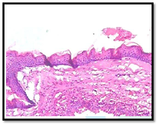
Microphotograph showing the histopathological features of KCOT (X10)

Microphotograph showing the histopathological features of KCOT(X40)
Histological variants of OKC (based on lining and types of keratin produced)


DISCUSSION
Redesignation of the OKC as the KCOT by the World Health Organization (WHO 2005) is based on the well-known aggressive behaviour of this lesion, its histology and new information regarding its genetics. The decision is based on following factors.
1. Behaviour: KCOT is locally destructive, highly recurrent and have Aggressive behavior. It recurrence rates is 3-60%. Recurrence rates reduced when more meticulous surgical treatment is done. Its penetration into cancellous bone can be very extensive. It is part of NBCCS.
Growth and Growth measurement is like a benign tumor:
KCOT grows not by osmotic expansion like other odontogenic cysts, it growth is by epithelial proliferation of wall with infolding of epithelial lining. It has unremitting growth.
Markers in cyst fluid of KCOT: Soluable protein content of cyst fluid in OKC < other cysts. AgNORs not diagnostic of OKC or other odontogenic cyst or unicystic ameloblastoma.
2.Histopathology : Basal layer of KCOT budding into the connective tissue and mitotic figures are frequently found in suprabasal layers. (Ahlfors et al., 1984; Barnes et al., 2005)
Genetics/Immunohistochemistry: Expression of P53 gene protein is strongly associated with aggressive behavior and high recurrence of KCOT (Kichi et al., 2005).
The neoplastic potential of odontogenic keratocyst (OKC) was further substantiated by these findings which suggests an alteration in gene expression. (Shear, 2002)
Histochemical and molecular studies shows that there is higher expression of oxidative enzymes (NADH2-, NADPH2-, G6PD and acid phosphatase) in OKC when compared to radicular, residual and dentigerous cysts. Leucine aminopeptidase is greater in OKC attributed to invasiveness of lesion.
Parathyroid hormone related protein PTHrP higher levels in OKC as compared to residual and dentigerous cysts which is related to bone resorption.
Again WHO has categorized OKC into Odontogenic and non-odontogenic developmental cysts (2017). It is no longer considered a neoplasm as the evidence supporting that hypothesis like clonality is considered insufficient.1
RECURRENCE OF OKC/KCOT
In 1976, Brannon proposed mechanisms for KCOT recurrence: incomplete removal of the cyst lining, growth of a new KCOT from satellite cysts (or odontogenic rests left behind after surgery) and development of a new KCOT in an adjacent area that is interpreted as a recurrence. It has high recurrence rate ranging from 25-60%1. Recurrence rates reduced when more meticulous surgical treatment is done. Majority of cases reported recurrence within 5 years post treatment. The mean time of recurrence for males was found to be 4years and for females it came out to be 7 years.
The causes and factors responsible for KCOT recurrence are :
1. Incomplete removal of cystic lining
2. Thin and friable nature of epithelial lining,
3. Higher level of cell proliferative activity in the epithelium.
4. Budding in the basal layer of the epithelium
5. Bony perforation.
6. Adherence to adjacent soft tissue.
7. Supraepithelial and Subepithelial split of the epithelial lining.
8. Parakeratinization of the surface layer
9. Remnants of dental lamina epithelium not associated with original OKC and development of new OKC in the adjacent area.
10. Growth of new OKC from satellite cyst /daughter cyst/remnants/cell rests.

(Madras J, Lapointe H. Keratocystic Odontogenic Tumour: Reclassification of the Odontogenic Keratocyst from Cyst to Tumour. J Can Dent Assoc. 2008 Mar;74(2): 165-165h )
CONCLUSION
There are several important things to remember about OKC.
- Unlike other jaw cysts this cyst has high recurrence rate estimated to be about 30%.
- Radiographically it is a great mimic. It may resemble periapical cyst, dentigerous cyst, lateral periodontal cyst, nasopalatine duct cyst, traumatic duct cyst and even tumors such as ameloblastoma.
- A person may have multiple keratocysts at the same time, a little unusual for jaw cysts.
- If a person has more than one OKC, that person must be investigated for nevoid basal cell carcinoma syndrome.
- Silently OKC extend anterio-posteriorly shows aggressive behavior like that of tumor , but again WHO categorized OKC into odontogenic and non-odontogenic developmental cyst. .
REFFERNCES
- Odontogenic keratocyst (okc) or journey of okc from cyst to tumor to cyst again : comprehensive review with recent updates on - deepak passi et al -2017.
- Keratocystic odontogenic tumor (parakeratinised oodontogenic keratocyst) - a review - usha Sharma et al -2016.
- Pathogenesis of keratocystic odontogenic tumor-a review - sunayana tendon et al - 2014
- Odontogenic keratocyst of maxilla involving the sinus – okc to be a cyst or a tumour? - dr. jyothi mahadesh et al - 2010.
- Keratocystic odontogenic tumour: reclassification of the odontogenic Keratocyst from cyst to tumour - jonathan madras-2008
- Maurette PE, Jorge J, de Moraes M. Conservative treatment protocol of odontogenic keratocyst: a preliminary study. J Oral Maxillofac Surg 2006
- Barnes L, Eveson JW, Reichart P, Sidransky D, editors. Pathology and genetics of head and neck tumours. Lyon: IARC Press; 2005. WHO classification of tumours series.
- Sapp JP, Eversole LR, Wysocki GP. Contemporary oral and maxillofacial pathology. 2nd ed. St. Louis: Mosby; 2004.
Disclaimer from Guident
The views and opinions expressed in articles published in Guident are solely those of the authors and do not necessarily reflect the views of the editors, the editorial board or the publisher, Ivory India. Guident does not endorse any product, technique or treatment described. Clinical content is intended for qualified dental and medical professionals and is not a substitute for individual clinical judgement. Guident, its editors and the publisher accept no liability for any consequence arising from the use of information published in the journal.
How to cite this article
Dr. Gopa Kumar R. Nair Dr. Vinay Mohan Dr. Praveen Kumar Department of Oral Medicine And Research Radiology. Odontogenic Keratocyst - Cyst / Tumor?. Guident. 2017 Oct;10(11). Available from: https://ivoryindia.in/articles/oral-medicine/odontogenic-keratocyst-cyst-tumor.html