Overview of Osteogenic Distraction for Treatment of Maxillary Constriction
Yenepoya Dental College, Magalore, India
INTRODUCTION
The theory of distraction was first published in 1905 by Codivilla1. The combined surgical and orthodontic treatment for maxillary expansion was introduced in 1938 for skeletally matured patients. The first successful use of distraction on the femur of a significantgroup of patients was published in 19902. In 1999 the first bone-borne distractor was introduced3. Maxillary expansion by means of distraction is a nowadays widely used treatment.
However, there is no consensus in the searched literature regarding the surgical technique,the type of distractor used (tooth-borne or bone-borne), the existence, cause and amount of relapse and whether or not overcorrection is necessary.
HISTORY
History of orthodontic treatment for maxillary constriction
Growth at the suture occurs through deposition of new bone at the sutural margin by the adjacent cellular layer. Toward the end of fetal life the cellular layers decrease in thickness, indicating that the rate of growth is slowing down, and the number of fibers in the intermediate layer uniting the capsular layers decreases. In a study of human sutures from birth to 18 years, Latham and Burstonconcluded that after about 2 of 3 years the sutures of the skull in general functioned primarily as sites of union of bones, but localized remodeling is a continuing process.
Cranial sutures are unified before complete eruption of the third molar. Soon after this, facial sutures close, and the sutures connecting the cranial and facial complexes are the last to close4. Regarding the facial sutures, Sicher5 states that the closure of sutures in human beings starts, as a rule, in the middle 30s at the posterior end of the median palatine suture but that some facial sutures, including the frontozygomatic, may remain open even in older age groups. This view is supported by Wright6, who claimed the intermaxillary and palatine sutures to be unossified and susceptible to comparatively easy separation at as late an age as 35 years.
A conflicting view is expressed by Persson7, who found evidence of bony union at 17 years in the midpalatal suture. Latham and Burston8, however, found no evidence of synostosis in the same suture by the age of 18 years. An over-all view is expressed by Scott9, who believes that, although most facial sutures appear open on the surface of old skulls, some degree of union may be present in the substance of the suture. It is obvious therefore, that the available literature is inconclusive and conflicting. In clinical practice, skeletal correction of the transverse discrepancy via orthodontics (orthopedics) is successful until the age of approximately 14-15 years dependingon the gender of the patient. After this age, orthodontic widening becomes virtually impossible and very painful10,11,12. In general, it is assumed that closure of the midpalatalsuture prevents this type of expansion10,12.In the first part of nineteenth century, Lefoulon13,14 and Talma15 reported on maxillary expansion with a palatal or buccal C-shaped spring. A method, reserved for less severe cases, consisted of lateral thumb pressure, 'every morning and even many times daily', by the parent or the child itself. The first documented case of orthodontic correction of maxillary width discrepancies was by Angell16. He performed rapid maxillary expansion with the use of a jackscrew appliance in a 14-year-old girl. He observed that by turning the jackscrew daily, he was able to open the maxillary suture sufficiently in a period of 2 weeks. Angell16 mentions correction of maxillary width discrepancies by opening the midpalatal suture. In 1913, Schröder-Benseler17 presented the still-popular all-wire frame with a non-spring-loaded jackscrew, the hygienic appliance. Derichsweiler16 uses bonds to the premolar and molar, which are embedded into a split acrylic base plate with an incorporated conventional orthodontic expansion screw. In1961 Haas 'Reintroduced' rapid maxillary expansion (RME) and mentions in 1970 that the use of RME is ideally during the growth spurt18,19. Reichenbach& Brückl20published an excellent survey on orthodontic treatment of maxillary transverse hypoplasia in 1967.
History of surgical treatment for maxillary constriction
Once skeletal maturity has been reached, orthodontic treatment alone cannot provide a stable widening of the constricted maxilla in cases of deficiencies of more than 5 mm. In general, an orthodontist can camouflage transverse discrepancies less than 5 mm with orthopedic forces alone21. The literature mentions several problems accompanied by RME on mature patients,such as failure and or relapse and periodontal problems with the tooth-borneappliances22. Timms& Vero,sup>23 mention that 33-50% of the expansion has relapsed before stability is achieved. Others report the lack of movement of the maxillary halves; excessive tipping of the anchor teeth; buccal root resorption of the anchor teeth or even periodontal defects as the teeth are pushed though the buccal cortical plate, which lead to bony defects and gingival recession; unequal expansion and unpredictable relapse and the sensation of pain and necrosis of oral mucosa under the appliance. Bell and Starnbach24,25,26 report that activation of an appliance against mature sutures can lead to the sensation of pain and necrosis of oral mucosa under the appliance. These forces can also result in periodontal defects as the teeth are pushed though the buccal cortical plate, which lead to bony defects and gingival recession. These complications can be avoided by surgically releasing the osseous structures that resist the expansive forces24,26. Therefore the combination of surgical and orthodontic treatment is advocated for widening of the maxilla in skeletally matured patients. Advantages of SARME include improvement of periodontal health; improved nasal air flow; elimination of the negative space, which results in less visible tooth and gingival structures upon smiling27. There is also a cosmetic improvement of the buccal hollowing secondary to post-expansion prominence at the site of the lateral wall osteotomy24,26. Tooth extractions for alignment of dental arches are often unnecessary21.Brown28 probably first described a technique of SARME with midpalatal splitting in his textbook. Heiss25 probably first inaugurated the midline splitting in the anterior maxilla for the extension of the compressed maxillary arch for orthodontic reasons. In 1961, Haas19 described the downward and forward movement of the maxilla that occurs during RME because of the location of the Cranio Maxillofacial sutures. He believed that the maxillary halves separated from each other rather in a tipping than in aparallel fashion due to the strength of the zygomatic buttresses19. Isaacson & Ingram29and Isaacson et al.30 mention that historically, the midpalatal suture was thought to be the area of resistance to expansion, but the facial skeleton increases its resistance to expansion as it ages and matures, and that the major site of resistance is not the midpalatalsuture but the remaining maxillary articulations. Wertz31 advocated that resistance of the zygomatic arch prevents parallel opening of the midpalatal suture. In 1975, Lines32 and in 1976 Bell & Epker24 demonstrated that the area of increased facial skeletal resistance to expansion was indeed not the midpalatal suture, but the zygomaticotemporal, zygomaticofrontal and zygomaticomaxillary sutures. Identification of these areas of resistance in the craniofacial skeleton stimulated the development of various maxillary osteotomies to expand the maxilla laterally in conjunction with orthodontic RME appliances4. The areas of resistance to lateral forces in the midface are the piriform aperture (anterior), the zygomatic buttress (lateral), the pterygoid junction (posterior) and the midpalatalsynostosed suture (median). In the early reports all four are transsected25,33,34,35 . In 1972 Steinhauser36 reportsa maxillary expansion osteotomy technique without the use of distraction, a Le Fort I type of osteotomy in combination with the surgical splitting of the palate in the midline, after which a triangular unicorticaliliac graft is inserted into the void created by the expansion. More recently, with the emphasis on decreased morbidity and ambulatory surgery, fewer supports are osteotomized; the anterior, lateral and median, the lateral and median,the anterior, posterior and lateral, the anterior and lateral. Most reports note that surgically assisted maxillary expansion is more stable than orthodontic RME alone24,34,35,37.
Glassmann et al.38, Alpern& Yurosko39 and Lehmann & Haas37 reported successful expansion in humans performed with a Hyrax appliance following a lateral osteotomy from the piriform rim to the pterygoid plate without palatal surgery. Their studydid not consider the amount of skeletal versus dental expansion and the corresponding relapse following a retention period40. In 1984 Glassmann et al. postulates that uniform palatal expansion can be achieved without sectioning of either palate or the pterygomaxillary fissure38.
In the year 1999, Mommaerts45 presented the Trans Palatal Distractor (TPD),which is a bone-borne device for SARME. After surgical release of the areas of maxillary support the tooth-borne devices used for SARME cause undesired movements of the abutment teeth during expansion and retention phases that could lead to periodontal problems35, 38, 41. Prolonged retention and overcorrection is advisable to counteract skeletal relapse. The TPD avoids all of these aforementioned problems, since fixation is sought in palatal bone32. Recently, the Magdenburg Palatal Distractor (PD) was presented, also a bone-borne device which claims to have no relapse42 .
HISTORY OF DISTRACTION
As mentioned before SARME is a form of distraction that was applied before its biological healing principles were known. Codivilla1 was the first to describe the technique of distraction osteogenesis for the shortened femur in 1905. Ilizarov described the use of distraction osteogenesis in the field of Orthopedics to lengthen the leg bones in a large group of patients in 19902. The technique is based on a 5-day period of rest after corticotomy before the expansion starts. This gives the tissue time to form the first callus but is too short for consolidation. Four phases of new bone formation can be described. The first is a fibrovascularheamatoma; between day 5 and 7 collagen fibers are formed that will arrange parallel to the distraction vector. Second,the bone formation follows the collagen fibers through intramembranous ossification; from the outside to the inside. Third, remodeling phase of the new bone.Fourth, formation of solid compact bone with the same texture as the surrounding (old) bones. When the distraction is performed too fast, the collagen fibers might lose contact and there is no in growth of new bone, providing non- or mal-union. In cases of a too slow distraction premature consolidation can occur and the requested elongation cannot be reached.
SURGICAL TECHNIQUE
Since early in the 20th century various techniques have been developed for SARME. The main considerations have opposing interests. One side is a more invasive technique with maximal mobility of the maxillary halves for correction over larger distances with less force but with more possible complications. The other side is less invasive with less possible complications but with more relapse, more periodontal problems, and unexpected fractures. The opinions vary about the site of major resistance in transverse distraction in the midface and also about the method of releasing it. Most methods consider the zygomaticomaxillaryjunction the major site of resistance and perform a corticotomythrough the zygomatic buttress from the piriform rim to the maxillopterygoid junction (fig 1)
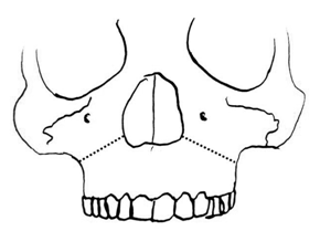
Figure 1. Schematic drawing showing the corticotomy from the piriform rim to the maxillopterygoid junction.
The midpalatal suture is historically considered the major place of resistance but this was proven to be untrue by Isaacson & Ingram29, Isaacson et al.30 and Kennedy et al.34 (Fig. 2). Still many, but not all, release the midpalatal suture to improve mobility and to prevent deviation of the nasal septum.

Figure 2.Schematic drawing showing the osteotomy of the midpalatal suture.
Several authors describe two paramedian palatal osteotomies from the posterior nasal spine to a point just posteriorly of the incisive canal (Fig. 3)9,11,57.

Figure 3.Schematic drawing showing the two paramedian palatal osteotomies from the posterior nasal spine to a point just posteriorly of the incisive canal.
The pterygoid plates are also a considerable site of resistance but because of the increased risk of injuring the pterygoid plexus by the osteotomy, some chose not to, without losing much mobility (Fig. 4). By not releasing the pterygoid junction, the pattern of opening of the maxillary halves is more V-shaped with the point of the V dorsally and it might be considered as an individual treatment to achieve more distraction either on the posterior or anterior level.

Figure 4.Schematic drawing showing the osteotomy of the pterygoid plates.
The nasal septum is often released from its palatal base to avoid shifting to either side and thereby causing changes in nasal flow (Fig. 5). A tomographic study by Schwarz showed no significant change in nasal septum position in SARME without sectioning of the nasal septum and an increase nasal airway space60.

Figure 5.Schematic drawing showing the release of the nasal septum with the use of a septum osteotome.
Of the studies on SARME mentioned in international literature, the mean age of the patients undergoing SARME varied from 19 to 29 years33,35,38,40,41,43,44,. The groups studied were quite small and mostly contained not more than 20 patients. The period of retention after expansion varies from 2 to 12 months. Generally, a period of three month is used. The amount of distraction at the canine level mentioned varies from 3.4 mm to 5.0 mm, in the first premolar region 4.7 mm to 5.9 mm and in the first molar region 3.4 mm to 8.0 mm. SARME is considered a procedure with little risk of serious complications , however several complications are mentioned in literature varying from life threatening epistaxis to a cerebrovascular accident, skullbase fracture with reversible oculomotor nerve pareses and orbital compartment syndrome12,35,. Less serious complications reported are postoperative hemorrhage, pain, sinusitis, palatal tissue irritation/ulceration, asymmetrical expansion, nasal septum deviation, periodontal problems and relapse46.
ACKNOWLEDGEMENT
The authors would like to acknowledge the contribution of Dr. Prakyath Malli in the review of this article
REFERENCES
1. Codivilla, A. On the means of lengthening, in the lower limbs, the muscles and tissues which are shortened through deformity. Am J OrthopSurg 1905: 2: 353.
2.Ilizarov GA. Clinical applications of the tension-stress effect for limb lengthening. ClinOrthop 1990: 250: 8-26.
3. Mommaerts MY. Transpalatal distraction as a method of maxillary expansion. Brit J Oral MaxillofacSurg 1999: 37: 268-272.
4 Krogman WM. Studies in growth changes in the skull and face of anthropoids: Ectocranial and endocranial suture closure in anthropoids and old world apes. Am J PhysAnthropol 1930: 46: 315-353.
5. Sicher H. Oral anatomy. ed. 4, St. Louis, 1965, the C.V. Mosby Company: 71-75
6 ZahlChr, Gerlach KL. PalatinaldistraktorEininnovativeransatzfür die gaumennahterweiterung. Mund Kiefer GesichtsChir 2002: 6: 446-449.
7 Persson M. Structure and growth of facial sutures.Odontol Revy 1973: 24(6).
8. Latham RA, Burston WR. The postnatal pattern of growth at sutures of the human skull. Dent Pract 1966: 17: 61-67.
9. Scott JH. Dentofacial development and growth.oxford, 1967, Pergamon Press: 89-93
10. Melsen B. Palatal growth studied on human autopsy material. Am J Orthod 1975: 68: 42-54.
11. Öztürk M, Doruk C, Özeç I, Polat S, Babacan H, Biçakci A. Pulpal bloodflow: effects of corticotomy and midline osteotomy in surgically assisted rapid palatal expansion. J Cranio- Maxillofacial Surg 2003: 31: 97-100.
12. Pogrel MA, Kaban LB, Vargervik K, Baumrind S. Surgically assisted rapid maxillary expansion in adults. Int J Adult OrthodonOrthognath Surg. 1992: 7: 37-41
13. Lefoulon PJ. Orthopédiedentaire.Gazette de l’hôpital. 1839: 111-119.
14. Lefoulon PJ. Nouveau traitéThéoriqueetpratique de l’artdentaire du dentiste. Pairs: fortin, Masson. 1841: 198.
15 Talma AF.Mémoires sur quelques points fondamentaux de la medicine dentaire. Bruxelles: Tircher. 1852.
16. Angell EH. Treatment of irregularity of the permanent or adult teeth. Dental Cosmos 1860: 1: 540-544, 599-600.
17. Schröder-Benseler. Die kiefererweiterung. Erg gesZahnhk.1913: 629.
18. Haas AJ. Rapid Palatal expansion of the maxillary dental arch and nasal cavity by opening the midpalatal suture. Angle Orthod 1961: 31: 73-90.
19. Haas AJ, Palatal expansion: Just the beginning of dentofacial expansion. Am J Orthod 1970: 57: 219-255.
20. Reichenbach E, Brückl H. Zahnärztlichefortbildung heft 7; Kieferorthopädischeklinik und therapie. Johann Ambrosius Barth, Verlag, Leipzig. P. 101.
21. Silverstein K, Quinn PD. Surgically-assisted rapid palatal expansion for management of transverse maxillary deficiency. J Oral MaxillofacSurg 1997: 55:725-727.
22. Pinto PX, Mommaerts MY, Wreakes G, Jacobs WVGJA. Immediate postexpansion changes following the use of the transpalatal distractor. J Oral MaxillofacSurg 2001: 59: 994-1000.
23. Timms DJ, Vero D. The relationship of rapid maxillary expansion to surgery with special reference to midpalatalsynostosis. Br J Oral Surg 1981: 19: 180-196.
24. Bell WH, Epker BN. Surgical orthodontic expansion of the maxilla. 1976: 70: 517-528.
25. Bell RA. A review of maxillary expansion in relation to rate of expansion and patients age. Am J Orthod 1982: 81: 32-37.
26. Starnbach H, Bayne D, Cleall J, Subtelny JD. Facioskeletal and dental changes resulting from rapid maxillary expansion. Angle Orthod 1966: 36: 152-164
27. Swennen G, Schliephake H, Dempf R, Schierle H, Malevez C. Craniofacial distraction osteogenesis: a review of the literature. Part I: clinical studies. Int J Oral MaxillofacSurg 2001: 30: 89-103
28. Brown GVI. The surgery of oral and facial diseases and malformation. 1938. 4th Ed., London; Kimpton: p.507.
29. Isaacson RJ, Ingram AH. Forces produced by rapid maxillary expansion I: Forces present during treatment. Angle Orthod 1964: 34: 256-260.
30 .Isaacson RJ, Wood JL, Ingram AH. Forces produced by rapid maxillary expansion II. Angle Orthod 1964: 34: 261.
31. Wertz RA. Skeletal and dental changes accompanying rapid midpalatal suture opening. Am J Orthod 1970: 58: 41-66.
32. Lines PA. Adult rapid maxillary expansion with corticotomy. Am J Orthod 1975: 67: 44-56.
33. Bell WH, Jacobs JD. Surgical-orthodontic correction of horizontal maxillary deficiency. J Oral Surg. 1979: 37: 897-902.
34. Kennedy JW, Bell WH, Kimbrough OL, James WB.Osteotomy as an adjunct to rapid maxillary expansion. Am J Orthod 1976: 70: 123-137.
35 .Kraut RA.Surgically assisted rapid maxillary expansion by opening the midpalatal suture. J Oral MaxillofacSurg 1984: 42: 651-655.
36. Steinhauser EW. Midline splitting of the maxilla for correction of malocclusion. J Oral Surgery 1972: 30: 413-422.
37. Lehman JA, Haas AJ. Surgical-orthodontic correction of transverse maxillary deficiency.ClinPlastSurg 1989: 16: 749-755.
38. Glassman AS, Nahigian SJ, Medway JM, Aronowitz HI. Conservative surgical orthodontic adult rapid palatal expansion: sixteen cases. Am J Orthod 1984: 86: 207-213.
39. Alpern MC, Yurosko JJ. Rapid palatal expansion in adults. Angle Orthod 1987: 57: 245-263.
40. Mossaz CF, Byloff FK, Richter M. Unilateral and bilateral corticotomies for correction of maxillary transverse discrepancies. Eur J Orthod 1992: 14: 110-116.
41. Bays RA, Greco JM. Surgically assisted rapid palatal expansion: an outpatient technique with long-term stability. J Oral MaxillofacSurg 1992: 50: 110-113.
42. Gerlach KL, Zahl C. Transversal palatal expansion using a palatal distractor. J OrofacOrthop 2003: 64: 443-449.
43. Chung CH, Woo A, Zagarinsky J, Vanarsdall RL, Fonseca RJ. Maxillary sagittal and vertical displacement induced by surgically assisted rapid palatal expansion. Am J OrthodDentofacialOrthop 2001: 120: 144-148.
44. Neubert J, Somsiri S, Howaldt HP, Bitter K. Die operative gaumennahterweiterungdurcheinemodifizierte Le-Fort-I-osteotomie. Dtsch Z Mund Kiefer GesichtsChir 1989: 13: 57-64.
45. Strömberg C, Holm J. Surgically assisted, rapid maxillary expansion in adults. A retrospective long-term follow-up study. J Cranio Maxillofacial Surg 1995: 23: 222-227.
46. Mehra P, Cottrell DA, Caiazzo A, Lincoln R. Life-threatening, delayed epistaxis after surgically assisted rapid palatal expansion: a case report. J Oral Maxillofac Surg. 1999: 57: 201-204.
Disclaimer from Guident
The views and opinions expressed in articles published in Guident are solely those of the authors and do not necessarily reflect the views of the editors, the editorial board or the publisher, Ivory India. Guident does not endorse any product, technique or treatment described. Clinical content is intended for qualified dental and medical professionals and is not a substitute for individual clinical judgement. Guident, its editors and the publisher accept no liability for any consequence arising from the use of information published in the journal.
How to cite this article
Dr. Faizan Ahmad Khan Dr. Anthony Kevin Fernandes Dr. Muraleedhara Bhat Department of Orthodontics and Dentofacial Orthopedics. Overview of Osteogenic Distraction for Treatment of Maxillary Constriction. Guident. 2017 Oct;10(11). Available from: https://ivoryindia.in/articles/orthodontics/overview-of-osteogenic-distraction-for-treatment-of-maxillary-constriction.html