Evaluation of the Effect of Dentin Desensitizers on the Retention of Full Metal Crowns Using Three Different Luting Cements
Solapur, Maharashtra
ABSTRACT
Frequently patients exposed to fixed restorative procedures experience discomfort in the prepared teeth either during the treatment or sometimes after cementation of restoration, which they perceive in the form of sensitivity due to which the clinical application of desensitizing agents has become prevalent. This study investigated the effect of GLUMA desensitizer and Bis Block on the retention of full metal crowns when cemented with three luting agents namely zinc phosphate, glass ionomer & resin cement .This study also evaluated the mode of action of desensitizers with Scanning Electron Microscopic (SEM) analysis and the nature of bond failure of the luting agents.
Keywords: desensitizing agents, retention, Scanning Electron Microscopic (SEM) analysis, smear layer, microleakage.
INTRODUCTION
Dentin reduction and exposure of prepared tooth surface can lead to increased dentin permeability and subsequent pulpal irritation in the form of sensitivity1. Dentin can become sensitive as a result of interim restoration microleakage and the resultant formation of bacterial byproducts2. Hypersensitivity has been reported even after cementation of definitive prostheses with the most widely used luting agents like zinc phosphate and glass ionomer cement due to the acidic nature of these cements which results in a partial removal of the smear layer3. The cement can enter the dentinal tubules before the luting agent sets displacing an equal volume of dentinal fluid in the pulp, which may cause excessive hydrostatic pressure leading to post-cementation sensitivity. However, with the use of a thin layer of dentin desensitizing agent, tubules can remain blocked, and the effect of external agents on dentin sensitivity can be greatly reduced. Application of desensitizing agents is gaining popularity. This study examined the effect of two commercially available desensitizing agents, non-polymerizable (GLUMA Desensitizer, Heraeus Kulzer, Germany) and polymerizable (BisBlock, Bisco, USA ), on the retention of complete veneer cast crowns when cemented with three luting agents namely zinc phosphate (Harvard cement, Germany) , Glass ionomer (Fuji I, GC Corp, Japan) and Resin cement (C&B Cement, Bisco ,USA). This study also evaluated the manner in which the desensitizers seal the dentinal tubules with Scanning Electron Microscopic (SEM) analysis and the nature of bond failure of the luting agents.
MATERIAL AND METHODS
Ninety freshly extracted human molars (of similar size) were selected for the study, cleaned and stored in distilled water at room temperature, with 0.1% thymol as preservative. They were then mounted in metal mould with selfpolymerizing acrylic resin and were stored in distilled water. A metal jig was prepared for holding the specimens at the time of tooth preparation. Standardized tooth preparation of 6 degrees total convergence, 4 mm axial height was done on each tooth with a custom made, air-rotor hand piece holding device that was attached to dental surveyor (Fig.1 and Fig.2). Occlusal and axial reduction were done by using wheel shaped diamond (WR- 13, Dia-Burs, Mani Inc. Japan) and the long round ended tapered diamond instrument (TR-13, Dia - Burs, Mani Inc, Japan) respectively. Finishing of the preparations was then done by using extra fine grit (20-30µ) round ended tapered diamond abrasive (TR-13EF, Dia-Burs, Mani Inc, Japan). A uniform 0.5mm chamfer gingival finish line, corresponding to half the diameter of the diamond point at its tip was prepared above the cementoenamel junction. The surface area of the preparations was measured, and was then categorized according to the descending order. The prepared specimens were divided into three groups and each group was further divided into three subgroups, so that each cementation group contained specimens of almost similar mean surface area. Impressions of the prepared teeth were made in addition silicone- putty & wash impression material (3M ESPE ExpressTM STD: 3M ESPE India Limited) by using metal rings of 25mm X 25mm size as special trays. The impressions were poured in type IV die stone (Ultrarock; Kalabhai Karson Pvt.Ltd, Mumbai, India). Wax coping of 0.5mm thickness and flat occlusal surfaces were made on dies with type II casting wax (Schuler – Dental – ULM, Germany). A loop was then attached onto the center of the occlusal surfaces of the pattern parallel to the long axis of the specimen. Individual castings were made from base metal alloy. Castings were finished, polished and checked for a good fit on the prepared teeth (Fig.3) and thereafter cleaned in an ultrasonic bath for 60 seconds.
Thirty samples (Group-G) were treated with GLUMA desensitizer and thirty samples (Group-B) were treated with BisBlock desensitizer. Thirty samples serving as control (Group-C) were left untreated. The ten crowns of each group were then cemented with Zinc phosphate (Subgroup C1, G1 and B1), Glass ionomer (Subgroup C2, G2 and B2) and Resin cement (SubgroupC3, G3 and B3). For achieving constant film thickness of the cement, a constant seating force was maintained during cementation. The crowns that were lined with cement were initially seated with strong thumb pressure. Thereafter, the assembled teeth and crown were placed on the customized jig attached to lower part of the Tinius Olsen universal testing machine and a static axial load of 5 kg for 10 minutes was applied (Fig.4). The excess cement was removed after setting of the cement using an explorer. After cementation, all the samples were stored at 370 C for 24 hrs before testing for retention. Then crowns were thermo-cycled between 5ºC and 55ºC water reservoirs for 600 cycles with a 28-second dwell time at each temperature. The retention testing of all the samples were performed on the Automated Universal Testing Machine (Fig.5). A self-alignment jig mounted on the universal testing machine was used for removal of crowns. The cross-head speed of the calibrated load cells was set at 1 mm/minute. Thus as the load cells moved apart at the designated crosshead speeds, a perfectly vertical tensional force was applied on the crowns and the tooth consistently until failure. The computer connected to the universal testing machine automatically recorded the loads and stress induced during testing. The maximum force at which the crown was separated from the tooth was recorded. The measured debonding force was divided by the tooth preparation area for each and converted into unit of stress (MPa). The data was organized and prepared for statistical analysis. Once the crowns had separated from the prepared teeth, the debonded surfaces were examined carefully to evaluate the mode of failure of the cement under magnification. The selected teeth were processed and scanned at desensitizer-dentin interface by using a Scanning Electron Microscope (S-3400N Analytical Scanning Electron Microscope). Areas showing characteristic features were located and photographed. (Fig.6, Fig.7, Fig.8).

Fig.1 Standardized tooth preparation using customized metal-zig
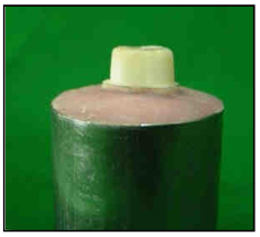
Fig 2 Prepare tooth
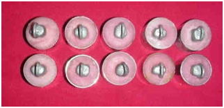
Fig 3 Casting fitted on the prepared teeth
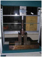
Fig.4 Cementation under constant load
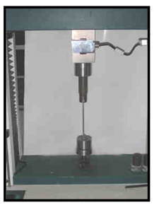
Fig 5: Tensile testing procedure
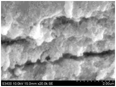
Fig 6 Scanning electron microscopy of control group
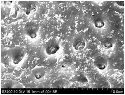
Fig7 Scanning electron microscopy of Bisblock group
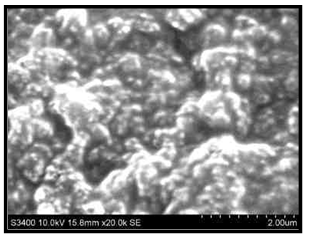
Fig.8 Scanning electron microscopy of Gluma desensitizer group
RESULTS
Table 1 (Master sheet) shows maximum stress values (in MPa), required to dislodge crowns. One way ANOVA test and multiple comparison test (Scheffe Analysis) was applied to all the groups. It was found that Resin cement produced the highest retentive strength values and all dentin treatments resulted in significantly different retentive strengths (p < 0.05): BisBlock > Control > Gluma desensitizer. The use of glass Ionomer cement resulted in retentive values between those of the resin and zinc Phosphate cement groups. None of the dentin treatment significantly affects the crown retentive strength of glass Ionomer cement (p Value > 0.05). Zinc Phosphate group demonstrated the lowest retention values and BisBlock treatment resulted in significant decrease in retentive strength compared with the Gluma desensitizer and Control groups.
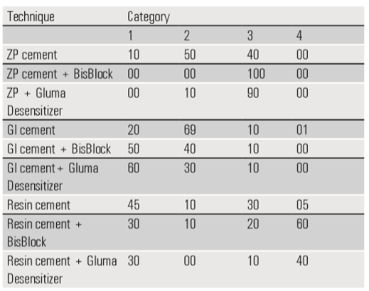
The results for characterization of failure mode are presented in Table 2 and Table 3. The modes of failure for castings cemented with zinc phosphate cement without desensitizer had cement principally on the castings and tooth. In contrast the cement was only found on the casting for the zinc phosphate group with desensitizers. With application of desensitizer, castings cemented with glass ionomer failed with cement generally residing on the preparation after crown removal, whereas control group demonstrated cement on both the preparation and casting. The resin cement without application of desensitizer predominantly remained on tooth surface, however with application of desensitizers predominant mode of failure was cohesive tooth fracture. Scanning Electron Microscopic Analysis of the Gluma desensitizer treated dentin revealed that the majority of tubules were obliterated with a thin layer that covered the surface and infiltrated into tubules as plugs. Bis-Block forms oxalate crystals deep within the dentinal tubules.
Table 1: Bond strength (in MPa) for groups of combination of cementing agents and desensitizers (n=10) (Master Sheet).
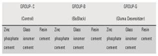
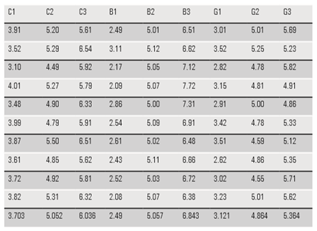
Table 2: Categories for characterization of type of failure after crown removal.
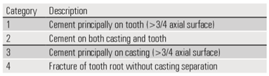
Table 3: Results showing the percentage of castings by failure type. The bolded values indicates the predominate failure modes.
DISCUSSION
The desensitizing agents applied on the tooth surface alter the retentive properties of the cements which ultimately alter the retentive strength of crown. In this study, in control group the mean retentive strength of zinc phosphate, glass ionomer and resin cement groups were 3.703 MPa, 5.052 MPa and 6.036 MPa, respectively
In the present study, the order of retentive strength of cements was resin cement > glass ionomer cement > zinc phosphate cement, despite of any desensitizing agent used. When glass ionomer cement was used with desensitizers retentive strength was BisBlock (5.057 MPa) > control (5.052MPa) > Gluma (4.864Mpa). However, the difference was not significant. Zinc phosphate had the lowest retentive values: control (3.703 MPa) > Gluma (3.121 Mpa) >BisBlock (2.49 Mpa). Yim NH et al concluded that Gluma desensitizers significantly reduces crown retention with zinc phosphate, glass ionomer and resin cement. The decrease in bond strength in all three groups was in correlation with the findings of the present study except the glass ionomer cement where the decrease in retentive strength was not significant. These findings confirm with Richardson D, Tao L, and Pashley DH4. They concluded that oxalate treated dentin did not affect the bond strength of glass-ionomer cement, but produced a large decrease in the bond strength of zinc phosphate cement. The present study also confirmed with the findings of Swift EJ et al5. They stated that Gluma desensitizer and One-step do not reduce the retention of cast metal crowns luted with glass ionomer cement.
SEM analysis in this study found that Gluma Desensitizer infiltrates into the tubules as plugs and form thin layer ovr the dentin occluding the entrance of the tubules similar to studies made by Arris CAG et al.6 SEM analysis in this study also shows calcium oxalate crystals in the opening of the dentinal tubules and small aggregates on the surfaces, confirming the results of studies by Pashley et al.,7; Tay et al.,8 of relieving post-operative sensitivity clinically is the adjunctive use of oxalate desensitizers on acid-etched dentin prior to adhesive application
Mode of failure of cementing agents supports the results regarding the variations in crown retentive values. With application of desensitizers, zinc phosphate cement predominantly remained on the castings which indicate that the retentive ability of the cement with tooth surface decreased drastically by application of desensitizing agents, especially with BisBlock. Glass Ionomer cement (with or without application of desensitizer) shows mixed type of failure with adhesive tendency more towards tooth. This supports the results with non-significant effect on crown retentive values. As far as resin cement is concerned most of the samples (40% with Gluma desensitizer, 60% with BisBlock) shows cohesive tooth failure without crown separation. This proves the results with significantly increased retentive values of resin cement.
CONCLUSION
Clinicians deal with crown and bridge restorative procedures on a regular basis. Desensitizing agents should be used routinely on all vital abutment teeth to be restored with fixed restorations not only to prevent dentin hypersensitivity but equally important to minimize bacterial contamination to reach the pulp. It is advantageous to use a polymerizable desensitizer when cementing casting with glass ionomer and resin cement, but it should not be used with zinc phosphate cement.
REFERENCES
- Richardson D, Tao L, Pashley DH. Dentin permeability; effect of crown preparation. Int J Prosthodont 1991; 4:219-25.
- Bergenholtz G. Inflammatory response of the dental pulp to bacterial irritation. J Endod. 1981; 7:100-4.
- Kern M, Kleimeier B, Schaller H, Strub JR. Clinical Comparison of postoperative sensitivity for a glass ionomer and a zinc phosphate luting cement. J Prosthet Dent. 1996;75:159-62.
- Richardson DW, Tao L, Pashley DH. Bond strengths of luting cements to potassium oxalate- treated dentin. J Prosthet Dent. 1990;63: 418-22.
- Swift EJ, Lloyd AH, Felton DA. The effect of resin desensitizing agents on crown retention. J Am Dent Assoc. 1997; 128:195-200.
- Arrais CAG, Chan DCN Giannini M. Effects of desensitizing agents on dentinal tubule occlusion. J Appl Oral Sci. 2004;12(2):144-8.
- Pashley DH, Carvalho RM, Pereira JC, Villanueva R, Tay FR: The use of oxalate to reduce dentin permeability under adhesive restorations. Am J dent. 2001;14:89- 94.
- Tay FR, Pashley DH, Mak YF, Carvalho RM, Lai SC, Suh BI. Integrating oxalate desensitizers with total etch two steps adhesive. J Dent Res. 2003b; 82: 703-707.
Disclaimer from Guident
The views and opinions expressed in articles published in Guident are solely those of the authors and do not necessarily reflect the views of the editors, the editorial board or the publisher, Ivory India. Guident does not endorse any product, technique or treatment described. Clinical content is intended for qualified dental and medical professionals and is not a substitute for individual clinical judgement. Guident, its editors and the publisher accept no liability for any consequence arising from the use of information published in the journal.
How to cite this article
Dr. Rajendra Avhad Goverment Medical College, Dr. Ranjukta Sar Dr.Jyoti Tembhurne Department of Prosthetic. Evaluation of the Effect of Dentin Desensitizers on the Retention of Full Metal Crowns Using Three Different Luting Cements. Guident. 2017 Oct;10(11). Available from: https://ivoryindia.in/articles/prosthodontics/evaluation-of-the-effect-of-dentin-desensitizers-on-the-retention-of-full-metal-crowns-using-three-different.html