Management of Anterior Open Bite in a Skeletal Class II Patient with Tongue Thrusting Habit: A Case Report
Department of Orthodontics and Dentofacial Orthopedics, A B Shetty Memorial Institute of Dental Sciences, Nitte (Deemed to be University), Mangalore, Karnataka, India
ABSTRACT
A 19-year-old male patient who was treated at the Department of Orthodontics and Dentofacial Orthopedics in A. B. Shetty Memorial Institute of Dental Sciences, Nitte (Deemed to be University) showed Angle’s Class II Subdivision malocclusion, skeletal class II relation due to mandibular retrusion, dental anterior open bite due to tongue thrusting and highly placed maxillary canines bilaterally. The diagnosis was established with the aid of a detailed case history and complete orthodontic records that included Extraoral/Intra Oral Examination, Model Analysis, and radiographic analysis.The treatment plan consisted of breaking the habit using the crib appliance, extraction of all 4 1st premolars, and closure of space with type A anchorage to correct the proclination of the anteriors.The fixed appliance used in this case was the 0.022” MBT appliance. Brackets were placed simultaneously in the upper and lower arches.
Objectives of the treatment were to:
- Intercept the Habit
- Align the upper and lower anteriors
- To bring the canine into the occlusal plane and to achieve Class I canine relation
- To have a functional Class I molar relation
- Correct the Midline Shift
- To Achieve Ideal Overjet and Overbite
INTRODUCTION
Caravelli gave the term “open bite” in 1842 to denote a distinct classification of malocclusion where there was a definite lack of contact in vertical dimention. Open bite may be anterior, posterior or combined. Anterior open bite (AOB) defined by Subtelny and Sakuda, is an open vertical dimension between the incisal edges of the maxillary and mandibular anterior teeth, although a deficiency in vertical dental contact can occur between the anterior or the buccal segment.1
Anterior open bite can be divided into two categories i.e., Dental; which results from dental eruption impediment and Skeletal; which is due to posterior facial growth or upward tipping of the maxillary plane.2 Dental AOB can lead to a negative impact on patients’ quality of life as it can affect their facial aesthetics and decrease their ability to communicate clearly. The characteristics seen are decreased incisor dentoalveolar height, normal or less anterior and posterior vertical dimensions, incompetent lips, and decreased incisal display at rest and smile.3 AOB has a multifactorial etiology and it has been associated with interactions among different factors, such as genetics, environment, oral habits, altered function/posture of tongue, neuromuscular deficiencies, and airway obstruction.4 The treatment for open bite problems ranges from observation or simple habit control procedures to complex surgical procedures. Extrusion of upper and lower incisor teeth, fixed orthodontics in conjunction with high?pull headgear therapy, temporary anchorage devices to intrude molar teeth, 2nd premolar extractions and multiple loop edgewise archwire therapy with vertical elastics are some treatment options.5
The case discussed here has a hyperdivergent growth pattern and retrognathic mandible with AOB and requires complex orthodontic treatments. The etiology appears to be environmental, which could be due to postural adjustments related to compromised airways and weak masticatory musculature. This leads to inadequate mandibular posturing.6 Such patients present with:
a) deficient ratio between posterior and anterior facial heights, giving rise to a long and convex facial profile
b) decreased masticatory function, with weak bite forces when compared to normal and hypodivergent subjects and
c) narrower dental arches, especially the maxillary one.
Hyperdivergent subjects present narrow maxillary dental arch when compared to normal and hypodivergent subjects. Also, any mandibular rotation present may be frequently camouflaged by remodeling of mandible, and only apparent rotation is clinically detected by orthodontists.7
Tongue thrusting habit is seen as a common cause of AOB. It occurs when the tongue makes contact with any of the teeth anterior to the molars during swallowing.Tongue thrust has been associated with posterior crossbite, open bite, and excess overjet in terms of malocclusion. Some common signs are hyperactive muscles i.e, mentalis, orbicularis oris and buccinator, and no momentary tooth contact when swallowing.8
The following case report illustrates the treatment of Angle’s Class II subdivision malocclusion with anterior open bite of 4.0 mm, a steep mandibular plane angle,and a Class II skeletal pattern further complicated with the presence of tongue thrusting habit.
CASE REPORT
DIAGNOSIS AND TREATMENT PLAN
A 19-year-old male patient came to the Department of Orthodontics and Dentofacial Orthopedics in A B Shetty Memorial Institute of Dental Sciences, Nitte (Deemed to be University) with a chief complaint of “Irregularly placed upper and lower teeth”
Patient-reported no relevant medical history that could impact the course of the treatment. Dental History showed a history of tongue thrusting since his adolescent years. The patient suggested that he was an ambivert and said that he was internally motivated to do the orthodontic treatment. He also said that the reason for undergoing orthodontic treatment was purely for esthetic imporvement.
Physical status examination showed that the patient had an athletic build with an unaltered gait, erect posture and a mesomorphic body type.
Upon facial analysis, the patient showed a Leptoprosopic facial type, a Dolicocephalic head typ,e a convex profile with posterior divergence. (Figures 1)
The mouth width was not coincident with the interpupillary distance. Neither was the nasal width coincident with the inner intercanthal distance. The patient had potentially competent lips with a low lip line.
Skeletally, the midlines were coincident. The patient had an average smile line and an acute nasio labial angle.

Figure 1: Pre Treatment Photographs. A – Frontal View (Incompetent Lips),B – Profile View (Potentially competent Lips), C – Smiling (Oblique View), D – Smiling (Frontal View)
Upon intraoral examination, an anterior open bite from lateral incisor to lateral incisors was observed. A dental midline shift was observed (Upper, 3 mm to the Left and Lower, 2 mm to the right). There was no overjet or overbite present. Unilateral Cross bite was present with respect to 22. The patient had a class II end on relation on the right side and Class I molar relation on the left side. Canine relation could not be assessed due to the highly placed canines. The patient had a V-Shaped maxilla and mandible with severe crowding in the upper anterior region and moderate crowding in the mandibular anterior region. The curve of spee was flat.
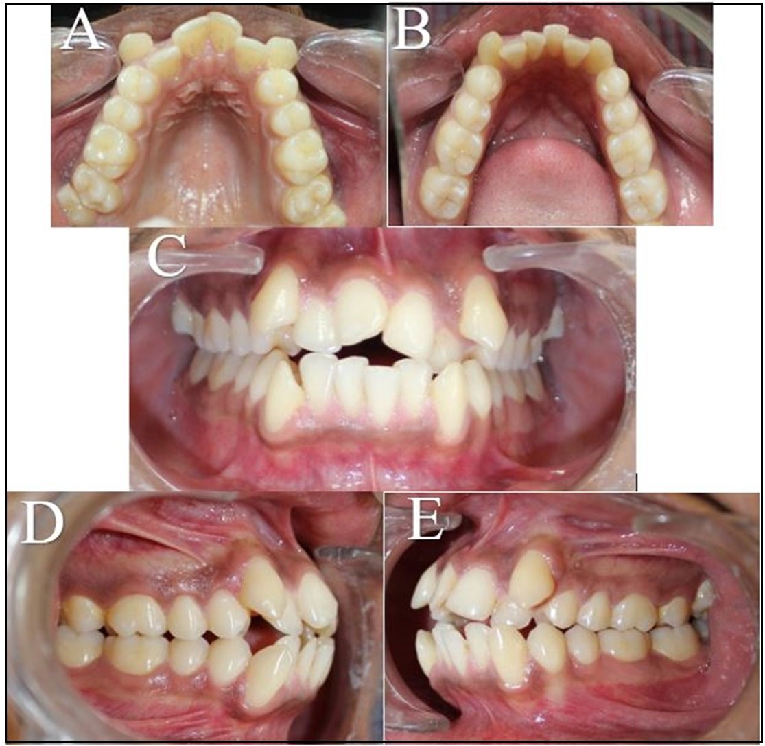
Figure 2: Pre Treatment Photographs. A – Maxillary Arch (Occlusal View), B –Mandibular Arch (Occlusal View), C – Frontal View, D – Right Side in Occlusion, E – Left Side in Occlusion
Radiographically, the panoramic radiograph evidenced the presence of all 4 third molars with the 48 being mesio-angularly impacted. On the lateral cephalogram a composite cephalometric analysis was performed (Table 1-3) which allowed us to diagnose the case as a skeletal class II due to mandibular retrusion. The analysis further revealed that the patient had a vertical growth pattern as well as upper and lower incisor proclination. Soft Tissue cephalometric values show that the patient has procumbent upper and lower lips
|
Skeletal Parameter |
Mean Value |
Standard Deviation |
Pre-Treatment Value |
Inference |
|
SNA |
81.77 |
6 |
78.6 |
Normal A-P Position of the maxilla |
|
SNB |
80.42 |
5.3 |
72.7 |
Retruded Mandible |
|
ANB |
2.05 |
1.8 |
5.83 |
Skeletal Class II |
|
Bjork Sum |
393.97 |
3.7 |
405.30 |
Hyperdivergent Skeletal Pattern |
|
FMA |
25 |
4.0 |
35.01 |
Hyperdivergent facial pattern |
|
Gonial Angle |
122.38 |
4.9 |
131.73 |
Obtuse Gonial angle |
|
APDI |
85.98 |
4 |
78.31 |
Skeletal Class II |
|
A to N Perp |
1.1 |
2.7 |
-1.21 |
Normal A-P Postion of the maxilla |
|
B to N Perp |
-3.5 |
2 |
-13.63 |
Retruded Mandible |
|
Pog to N Perp |
-0.3 |
2.5 |
-15.97 |
Retruded Chin |
|
Wits Appraisal |
-2.24 |
0.3 |
5.12 |
Skeletal Class II |
Table 1 - Pre Treatment Skeletal Cephalometric Values with Inference
|
Dental Parameter |
Mean Value |
Standard Deviation |
Pre-Treatment Value |
Inference |
|
Overjet |
2 |
2 |
0 |
Edge to Edge / Anterior Open Bite |
|
Overbite |
2 |
2 |
-3.70 |
Anterior Open Bite |
|
U1 to FH |
116.52 |
6 |
130.18 |
Proclined Upper Incisor |
|
U1 to SN |
109.31 |
6 |
119.89 |
Proclined Upper Incisor |
|
U1 to UOP |
55 |
4 |
44.94 |
Protruded Upper Incisor |
|
IMPA |
90 |
3.5 |
103.19 |
Proclined Lower Incisor |
|
L1 to LOP |
66 |
5 |
53.85 |
Protruded Lower Incisor |
|
Interincisal Angle |
128 |
5.3 |
91.62 |
Proclined Incsiors |
|
Cant of Occlusal Plane |
9.3 |
3.8 |
8.30 |
Normal Occlusal plane |
|
U1 to NA (mm) |
4 |
3 |
9.97 |
Protruded Upper Incisor |
|
U1 to NA (Deg) |
22 |
5 |
41.29 |
Proclined Upper Incisor |
|
L1 to NB (mm) |
4 |
2 |
15.81 |
Protruded Lower Incisor |
|
L1 to NB (Deg) |
25 |
5 |
41.26 |
Proclined Lower Incisor |
Table 2 - Pre Treatment Dental Cephalometric Values with Inference
|
Soft tissue Parameter |
Mean Value |
Standard Deviation |
Pre-Treatment Value |
Inference |
|
Upper Lip to E Plane |
0 |
2 |
1.72 |
Normal Upper Lip Position |
|
Lower Lip to E Plane |
0 |
2 |
8.69 |
Protruded Lower Lip |
|
Nasio Labial angle |
95 |
5 |
83.77 |
Protruded Lip |
Table 3 : Pre Treatment Soft Tissue Cephalometric Values with Inference
Model Analysis showed an Overall Boltons Ratio of 89.3% with overall maxillary excess of 2.2 mms and an Anterior Boltons Ratio of 76.9 with anterior maxillary excess of 0.2 mm
Carey’s and Arch Perimeter Analysis showed Tooth Material and Arch Perimeter discrepancy of 13mm in the upper arch and 9 mm in the lower arch suggesting 1st premolar extractions for the treatment.
Ashley Howe’s Analysis showed that expansion was needed in the upper and lower arch and that both the arches belonged to the ‘Definite Extraction’ category.
TREATMENT OBJECTIVES
The objectives of the treatment were to:
- Eliminate the Tongue Thrusting Habit
- Align the upper and lower anteriors
- Bring the canine into the occlusal plane and to achieve Class I canine relation
- Have a functional Class I molar relation
- Correct the Midline Shift
- Achieve Ideal Overjet and Overbite
ALTERNATIVE TREATMENT PLANS
- Surgical Orthodontics
- Fixed Orthodontics with the Use of Infrazygomatic Implants for Intruding the maxilla in the posterior region.
- Camoflague Treatment.
TREATMENT PLAN
- Interception of tongue thrusting habit by using the tongue crib habit-breaking appliance
- Extraction of all 1st premolars (Camoflague)
- Leveling and Aligning
- Intrusion of Posteriors and Extrusion of Anteriors
- Finishing and Detailing
TREATMENT PROGRESS
Prior to the initiation of the treatment, all 4 1st premolars were extracted.
Treatment was initiated with the placement of a banded tongue crib appliance to eliminate the tongue thrusting habit. Simultaneously the upper and lower arches were bonded using American Ortho ‘Empower’ brackets which are passive self-ligating brackets with the MBT Prescription (Figure 3). The sequence of wires used for alignment were 0.012 Niti, 0.014 Niti, 0.016 Niti, 0.018 Niti, 16x22 Niti and 17x25 Niti.

Figure 3: A – Maxillary Cast with Tongue Crib (Occlusal View), B – Image depicting Height of the Crib, C – Fixed Appliance with Banded Tongue Crib (Right View), D – Fixed Applaince with Banded Tongue Crib (Left View), E - Fixed Appliance with Banded Tongue Crib (Frontal View)
0.016 AJ Wilcock wire was placed in the upper arch and lower arch along with open coil spring to create space between 31/42 and 21/23. This was to help engage the wire into 41 and to create space to bring 22 into the arch. Once enough space was created, 0.014 niti was engaged into the tooth in question to bring it into the arch. (Figure 4)
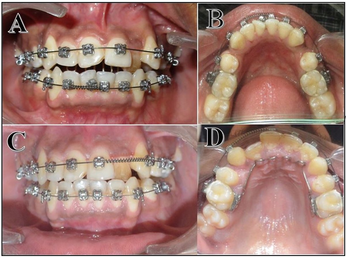
Figure 4: A – Open Coil Spring in lower arch to create space to engage 41. (Frontal View), B Open Coil Spring in lower arch to create space to engage 41. (Occlusal View), C – Open Coil Spring in the upper arch to create space for 22 (Frontal View), D – Open Coil Spring in the upper arch to create space for 22 (Occlusal View)
Once the leveling phase was completed, en-mass retraction of the six anterior teeth was begun with 0.017” x 0.025” stainless steel archwires and Elastomeric chain from the canine to the molar. Once Class I canine relation was achieved, the remaining spaces in the lower arch were closed by protraction of the posteriors.During space closure 3.5 Oz 3/16” class II intermaxillary elastics were used. (Figure 5)

Figure 5: A – Maxillary Arch after Retraction of Anteriors (Occlusal View), B –Mandibular Arch after Retraction of Anteriors (Occlusal View), C – Frontal View, D – Protraction of Posteriors using Open Coil Spring and Class II elastics (Right View), E – Protraction of Posteriors using Open Coil Spring and Class II elastics (Left View)
Once the spaces were closed a panoramic radiograph was obtained and the brackets of certain teeth were repositioned with the aim of obtaining root parallelism. Treatment continued with the settling phase by using a 5/16” 3½ Oz triangular elastic in the posterior region and 0.018 Niti archwires on the upper arch and lower arches Once occlusal settling was achieved, a fixed Lingual retainer was bonded in both arches just prior to appliance removal. (Figure 6) The fixed lingual retainer ran from 2 to 2 in the upper arch and 3 to 3 in the lower arch.
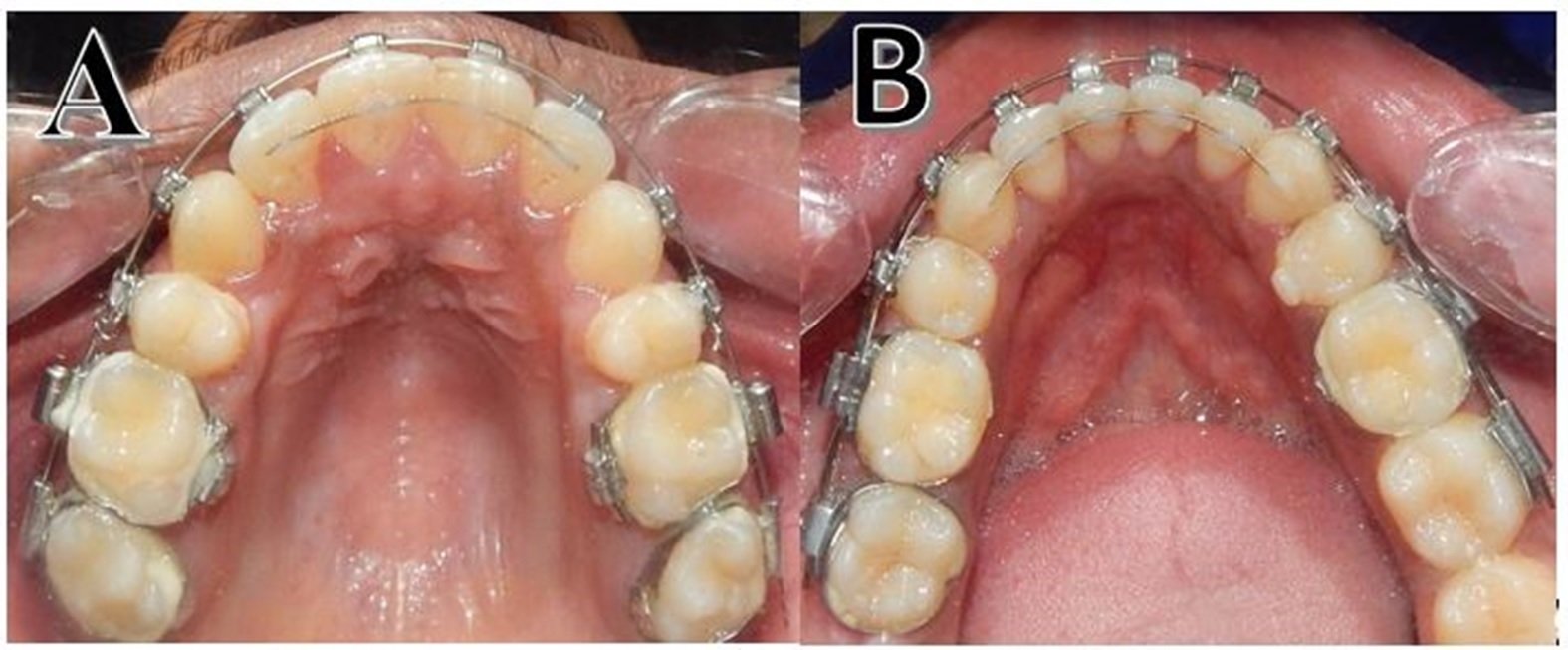
Figure 6 – A – Maxillary Arch with Bonded Lingual Retainer just prior to Debonding, B - Maxillary Arch with Bonded Lingual Retainer just prior to Debonding
Upon removal of the appliance, all post-treatment records were obtained (Figures 7,8) and a Hawleys Appliance was delivered to the patient.

Figure 7 - Post Treatment Photographs. A – Frontal View (Competent Lips),B – Profile View, C – Smiling (Oblique View), D – Smiling (Frontal View)

Figure 8 - Pre Treatment Photographs. A – Maxillary Arch (Occlusal View), B –Mandibular Arch (Occlusal View), C – Frontal View, D – Right Side in Occlusion, E – Left Side in Occlusion
The fixed treatment span was completed over a period of 18 months. The tongue crib therapy started simultaneously with the fixed treatment but ended 4 months prior to debonding the fixed appliance.
RESULTS
Lip competence was obtained as well as better lower lip projection and facial harmony which is evident when the Pre Rx and Post Rx are superimposed on each other.(Figure 9) Functional molar class II, canine class I and normal overjet were achieved.
Hard tissue changes were minimal.The obtained cephalometric changes were significant in the dental and soft tissue envelope through a decrease in protrusion and proclination. (Tables 4-6).
A point to be noted was that the worsening of the profile did not occur while retracting the teeth and this was probably because of good vertical control provided by the tongue crib appliance.

Figure 9 – Superimposition of Pre-Treatment Cephalogram (Black) and Post-Treatment Cephalogram
|
Skeletal Parameter |
Pre-Treatment Value |
Post Treatment Value |
|
SNA |
78.6 |
78.3 |
|
SNB |
72.7 |
73.2 |
|
ANB |
5.83 |
5.1 |
|
Bjork Sum |
405.30 |
405.07 |
|
FMA |
35.01 |
35.1 |
|
Gonial Angle |
131.73 |
131.19 |
|
APDI |
78.31 |
77.43 |
|
A to N Perp |
-1.21 |
-0.91 |
|
B to N Perp |
-13.63 |
-13.72 |
|
Pog to N Perp |
-15.97 |
-16.1 |
|
Wits Appraisal |
5.12 |
4.68 |
Table 4 : Comparison of Skeletal Cephalometric values between Pre Treatment and Post Treatment cephalogram tracing.
|
Dental Parameter |
Pre-Treatment Values |
Post-Treatment Values |
|
Overjet |
0 |
3 |
|
Overbite |
-3.70 |
1 |
|
U1 to FH |
130.18 |
120.98 |
|
U1 to SN |
119.89 |
111.11 |
|
U1 to UOP |
44.94 |
48.39 |
|
IMPA |
103.19 |
95.72 |
|
L1 to LOP |
53.85 |
57.26 |
|
Interincisal Angle |
91.62 |
108.10 |
|
Cant of Occlusal Plane |
8.30 |
9.52 |
|
U1 to NA (mm) |
9.97 |
7.51 |
|
U1 to NA (Deg) |
41.29 |
31.78 |
|
L1 to NB (mm) |
15.81 |
12.78 |
|
L1 to NB (Deg) |
41.26 |
34.03 |
Table 5 : Comparison of Dental Cephalometric values between Pre Treatment and Post Treatment cephalogram tracing.
|
Soft tissue Parameter |
Pre-Treatment Value |
Post-Treatment Values |
|
Upper Lip to E plane |
1.72 |
0.46 |
|
Lower Lip to E plane |
8.69 |
3.46 |
|
Nasio Labial Angle |
83.77 |
87.13 |
Table 6 : Comparison of Soft Tissue Cephalometric values between Pre Treatment and Post Treatment cephalogram tracing.
DISCUSSION
The best treatment plan for such a case i.e., Class II skeletal relation with a vertical growth pattern would have been surgical orthodontics. Differential Impaction with Posterior upward tipping of the maxilla and auto-rotation and advancement of the mandible would have solved both the sagittal and vertical discrepancies. Unfortunately, the patient was not willing to undergo surgery for the same. A great example of a Skeletal Class II case with anterior open bite being treated by Surgical orthodontics is shown by Miguel et al.9
The next best option would have been the use of Infrazygomatic Crestal implants for intruding the posterior aspect of the maxilla. While the patient did consent to this, due to the thin cortical bone present, none of the IZC’s were able to be successfully placed in his mouth. Arvind et al.10 have shown that hyperdivergent cases have the lowest density of bone when compared to normodivergent and hypodivergent subjects. This helps us understand why the IZCs could not be placed in this patient.
Therefore, camouflage treatment was the only option left for the aforementioned reasons. Most of the extraction space was utilized for the correction of crowding and slight retraction of the anteriors. The remaining space was used to protract the posteriors to achieve Class I molar relation.
CONCLUSION
All treatment objectives were completed satisfactorily. A significant change in the soft tissue profile was observed. Proclination of the upper and lower incisors were brought to near normal range, all within the limits of orthodontic tooth movement. Most importantly, the patient’s chief complaint was addressed, and the patient was satisfied with the results of the treatment.
REFERENCES
- Subtelny JD, Sakuda M. Open Bite: diagnosis and treatment. Am J Orthod 1964; 50: 337-358. Ref.: https://goo.gl/djgwjt
- Greenlee GM, Huang GJ, Chen SS, Chen J, Koepsell T, et.al. Stability of treatment for anterior open-bite malocclusion: a meta-analysis. Am J Orthod Dentofacial Orthop. 2011; 139: 154-169. Ref.: https://goo.gl/XuUaA1
- Islam ZU, Shaikh AJ, Fida M. Dentoalveolar heights in vertical and sagittal facial patterns. J Coll Physicians Surg Pak 2016;26:753?7., Nanda R. Biomechanics and Esthetic Strategies in Clinical Orthodontics. St. Louis, Mo. : Elsevier Saunders; c2005. p. 156?75
- L.H. Lin, G.W. Huang, C.S. Chen Etiology and treatment modalities of anterior open bite malocclusion J Exp Clin Med, 5 (2013), pp. 1-4
- Sbtelny J, Sakuda M. Open bite: Diagnosis and treatment. Am J OrthodDentofac Orthop 1964;50:337?58.
- Buschang PH, Jacob H, Carrillo R. The morphological characteristics, growth, and etiology of the hyperdivergent phenotype. InSeminars in Orthodontics 2013 Dec 1 (Vol. 19, No. 4, pp. 212-226). WB Saunders
- Proffit WR, Fields HW, Nixon WL. Occlusal forces in normal? and long?face adults. J Dent Res 1983;62:566?70
- Shetty S, Shaikh N. An effective and simple method for treatment of tongue thrusting in adult patient.
- Miguel JA, e Cal-Neto JP, da Silveira HM. Surgical correction of a Class II skeletal malocclusion associated with anterior open bite and temporomandibular joint pain. American Journal of Orthodontics and Dentofacial Orthopedics. 2007 Sep 1;132(3):400-7.
- Arvind TR P, Jain RK. Computed tomography assessment of maxillary bone density for orthodontic mini-implant placement with respect to vertical growth patterns. Journal of Orthodontics. 2021 Dec;48(4):392-402.
Disclaimer from Guident
The views and opinions expressed in articles published in Guident are solely those of the authors and do not necessarily reflect the views of the editors, the editorial board or the publisher, Ivory India. Guident does not endorse any product, technique or treatment described. Clinical content is intended for qualified dental and medical professionals and is not a substitute for individual clinical judgement. Guident, its editors and the publisher accept no liability for any consequence arising from the use of information published in the journal.
How to cite this article
Prof U S Krishna Nayak, Dr. Keerthan Shashidhar, Dr. Tarona Azem Subba. Management of Anterior Open Bite in a Skeletal Class II Patient with Tongue Thrusting Habit: A Case Report. Guident. 2022 Oct;15(11-12). Available from: https://ivoryindia.in/articles/orthodontics/management-of-anterior-open-bite-in-a-skeletal-class-ii-patient-with-tongue-thrusting-habit-a-case-report.html