Decompression and Enucleation of a Maxillary Radicular Cyst, Followed by Bone Regeneration, and Implant-Supported Rehabilitation of the Maxillary Anterior Region: A Case Report
Department of Prosthodontics and Crown & Bridge, Seema Dental College & Hospital, Rishikesh, Uttarakhand, India.
ABSTRACT
Immediate implanting has become a viable option to treat the oral rehabilitation process after a tooth loss by minimizing the number of surgical procedures, shortening the overall process, and increasing the comfort of the patient. However, implanting in areas involving periapical pathology, specifically those involving radicular cysts, is still a contentious issue because of the possible presence of infectious remains and a possible delay in the healing of the bone. The absence of sufficient clinical information regarding the predictability of using implants in previously infected areas keeps this issue a continuous debate in the field of implantology.
This case report presents a case in which a tooth associated with a radicular cyst was endosseously implanted. A 20-year-old male patient was diagnosed with a radicular cyst in the anterior maxilla. After the clinical and radiographic assessments, the decision was made for cystic lesion enucleation and immediate implant placement. Prior to the surgery, a cone beam computed tomography (CBCT) was done to evaluate the lesion and the bone around it. The surgery involved cystic lesion removal, infectious site debridement, implant placement to gain sufficient primary stability, and antibiotic prophylaxis.
The next day after the successful implant placement, a cement-retained provisional prosthesis was delivered. At the three-month follow-up, the patient had more positive than negative outcomes, demonstrating successful osseointegration with no peri-implant bone loss nor any recurrent pathology.
Keywords: Immediate Implant Placement, Radicular Cyst, Cortico-Basal Implant, Periapical Pathology, Oral Rehabilitation.
INTRODUCTION
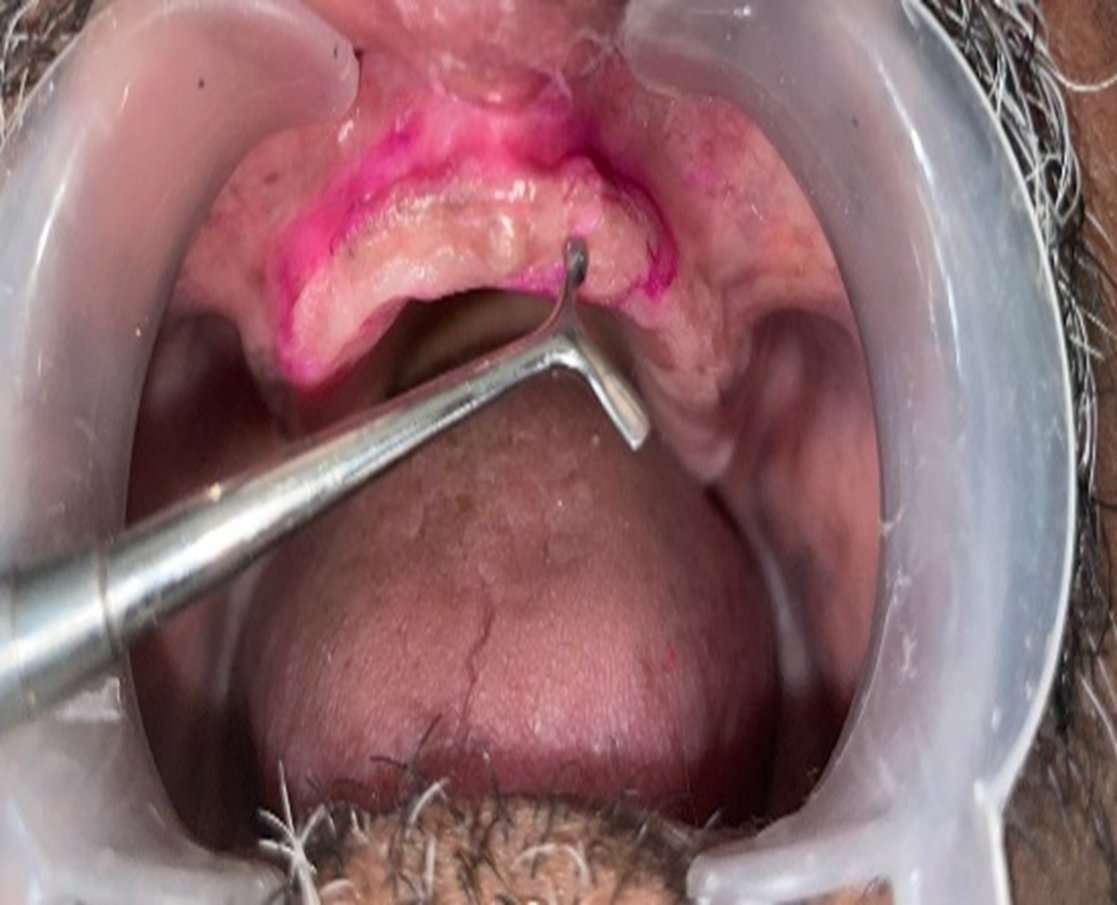
Childhood tooth loss is a frequent occurrence due to trauma. Avulsed maxillary anterior teeth can lead to long-lasting issues with function and even more to the patients’ esthetic concerns. Periapical pathologies can be quiescent for long periods and may be missed on routine.1 The clinical examination of the maxillary anterior segment revealed a partially edentulous area corresponding to the missing teeth #11, #12, and #21. The mucosa which was overlying the area was in a healthy state, and there were no signs of inflammation. Supra-eruption of the mandibular anterior teeth was noted. The patient was very concerned about the aesthetics of the area and opted for a fixed restorative option.
A Cone Beam Computed Tomography (CBCT) scan of teeth numbers 11, 12, and 21 showed a well-defined oval-shaped lesion that is osteolytic. The lesion measurement is 8.24 x 10.78 mm. The lesion is located in the area of the right central and lateral incisor. There were no signs of calcification in the radiolucent area of the lesion. The initial diagnosis is a radicular cyst based on the x-ray and clinical data.
A local anaesthetic was applied, and a crevicular incision was made in the area of teeth numbers 11, 12, and 21, and an envelope flap was raised. A full thickness mucoperiosteal flap was raised. Cystic lesion removal was done surgically, and the associated granulation tissue was removed using curettage. The area was rinsed with amoxicillin and irrigated to reduce the chance of bacterial infection.
Implant osteotomy was initiated using a pilot straight drill directed toward the palatal bone, and a paralleling pin was used to verify the angulation. This was followed by preparation with a 2 mm conical drill to create the initial osteotomy, after which the final osteotomy was completed using a 2.8 mm drill. After achieving satisfactory primary stability, two angulated implants were placed: a 3.75 × 13 mm implant in the #12 region and a 3.3 × 16 mm implant in the #21 region.
To manage the residual defect in the #11 and #12 areas, a xenograft bone substitute (Creos Xenogain, Nobel Biocare; 0.50 g vial) was placed over the grafted site. Primary wound closure was achieved using simple interrupted sutures. An immediate postoperative radiovisiograph (RVG) was taken to confirm the correct positioning of the implants. Postoperative management included a course of antibiotics and anti-inflammatory medications for one week. The patient was subsequently monitored through periodic follow-up visits for a duration of three months, during which no postoperative complications or adverse outcomes were observed.
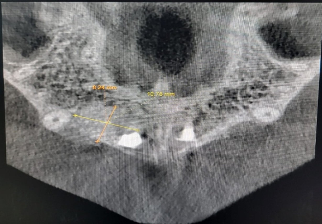
CBCT Scan showing anterior-posterior, transverse dimensions extension of the radicular cyst.
CASE REPORT
A 20-year-old male patient presented to the departement with concerns regarding the unaesthetic appearance of his maxillary anterior region and expressed a desire for a fixed prosthetic replacement. The patient reported a history of traumatic avulsion of teeth #11, #12, and #21 approximately 10 years earlier during childhood and had not undergone any prosthetic rehabilitation since the incident. He also complained of intermittent pain in the upper anterior region.
Clinical examination revealed a partially edentulous area in the maxillary anterior segment corresponding to the missing teeth #11, #12, and #21. The overlying mucosa appeared healthy without any visible signs of inflammation. Supra-eruption of the mandibular anterior teeth was also observed. The patient expressed significant concern regarding esthetics and requested a fixed restorative solution.
Cone Beam Computed Tomography (CBCT) of the region involving teeth #11, #12, and #21 was advised for further evaluation. The scan demonstrated a well-defined, oval-shaped osteolytic lesion measuring approximately 8.24 × 10.78 mm in the anteroposterior and transverse dimensions. The lesion extended in the region of the right central incisor and lateral incisor. Radiographically, the internal structure appeared completely radiolucent with no evidence of calcifications or internal septations. Based on the clinical findings and radiographic characteristics, a provisional diagnosis of radicular cyst was established.
Under local anesthesia, a crevicular incision was made in relation to the #11, #12, and #21 regions to raise an envelope flap, followed by elevation of a full-thickness mucoperiosteal flap. Surgical enucleation of the cystic lesion was performed, and the associated granulation tissue was thoroughly removed by curettage. The surgical site was irrigated with an amoxicillin solution to minimize bacterial contamination.
Implant osteotomy was initiated using a pilot straight drill directed toward the palatal bone, and a paralleling pin was used to verify the angulation. This was followed by preparation with a 2 mm conical drill to create the initial osteotomy, after which the final osteotomy was completed using a 2.8 mm drill. After achieving satisfactory primary stability, two angulated implants were placed: a 3.75 × 13 mm implant in the #12 region and a 3.3 × 16 mm implant in the #21 region.
To manage the residual defect in the #11 and #12 areas, a xenograft bone substitute (Creos Xenogain, Nobel Biocare; 0.50 g vial) was placed over the grafted site. Primary wound closure was achieved using simple interrupted sutures. An immediate postoperative radiovisiograph (RVG) was taken to confirm the correct positioning of the implants.
Postoperative management included a course of antibiotics and anti-inflammatory medications for one week. The patient was subsequently monitored through periodic follow-up visits for a duration of three months, during which no postoperative complications or adverse outcomes were observed.

Figure 1: Preoperative intraoral view showing missing maxillary anterior teeth (#11, #12, and #21) and compromised esthetics.

Figure 2: OPG images showing cystic lesion in the maxillary anterior region.

Figure 3: RVG showing the bone defect following cyst enucleation.

Figure 4: Placement of Noris Tuff implants in regions #12 and #21 with bone graft placed in the #11 region.
The patient was recalled after one week for suture removal. Mild swelling was observed due to graft seepage into the buccal soft tissue space; however, this subsided after steroid medication and subsequent follow-up visits. Graft seepage or displacement into the buccal soft tissue space is a recognized postoperative complication following bone grafting procedures and is frequently reported after guided bone regeneration and maxillary sinus floor augmentation procedures.3 In the present case, mild postoperative swelling was observed due to graft migration into the buccal region, which resolved following pharmacological management and close follow-up. Graft displacement is commonly associated with technical and surgical factors such as inadequate barrier membrane coverage4, membrane exposure, poor flap management, wound dehiscence5, insufficient condensation of graft material, excessive graft volume, and improper graft placement technique. Failure to create a stable and contained graft environment can allow particulate graft material to migrate into adjacent soft tissue spaces.6
Increased intraoral or intranasal pressure, such as during sneezing, nose blowing, or straining, can force graft particles out of the surgical site.7 Additionally, postoperative infection can compromise soft tissue integrity, leading to graft exposure and migration.8

Figure 5: Graft seepage in buccal vestibule

Figure 6: Second-stage surgery showing healing abutments in situ after 3 months of osseointegration.
PROSTHETIC PHASE
Supra-eruption of the mandibular anterior teeth posed a significant challenge in prosthetic rehabilitation. One Fifteen-degree and one straight abutment were used to correct implant angulation and improve prosthetic path of insertion. A digital impression was recorded, and an FP3-type prosthesis was fabricated to camouflage gingival contour discrepancies. The patient was satisfied with the esthetic and functional outcome. A water flosser was recommended to maintain hygiene around the implant-supported prosthesis.

Figure 7: Angled abutments and digital impression procedure

Figure 8: Final FP3 prosthesis – intraoral view.
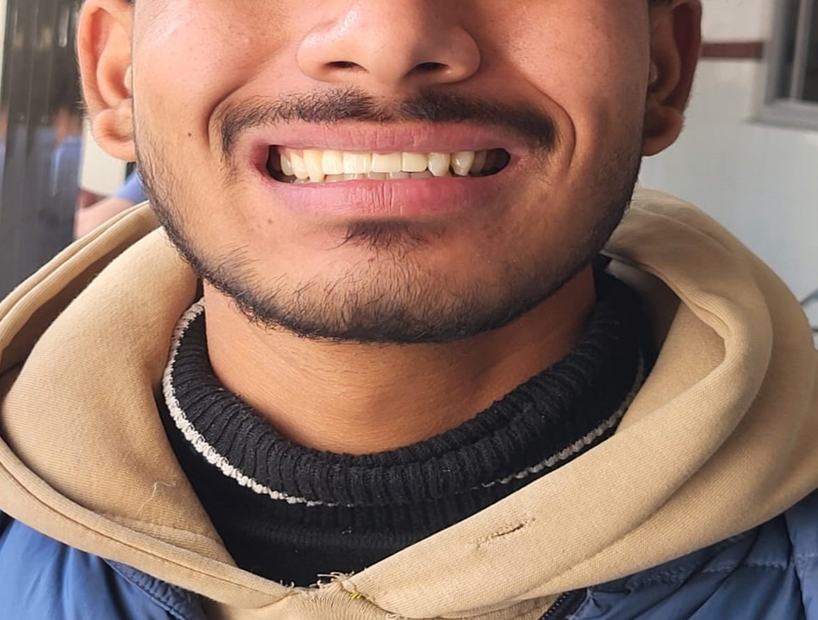
Figure 9: Post-treatment extraoral smile view
DISCUSSION
Considering that contamination might impede osseointegration and lead to loss of implants placed in sockets with periapical lesions. Periapical pathosis usually contraindicates their rapid replacement with endo-osseous dental implants.9 Extensive cystic lesions in the anterior maxilla can lead to significant alveolar bone destruction, posing challenges for implant placement due to compromised bone volume, altered anatomy, and difficulty in achieving primary stability.10,11 Early detection of such lesions is critical, as long-standing cysts can remain asymptomatic and may not be evident on conventional two-dimensional radiographs.12,13 Immediate implant placement provides benefits on both the social and economic fronts. It not only prevents the alveolar bone level from resorbing, but it also shortens the length of the treatment by combining tooth removal followed by implant placement along with or without bone grafting in single sitting, it reduces the number of surgical procedures.14
The use of angulated implants and abutments provides an increased antero-posterior (AP) spread, which is biomechanically advantageous in anterior fixed prostheses. Increased AP spread allows the prosthodontist to design cantilever extensions with favorable load distribution and reduced stress concentration on implants.15 Angulated abutments also help correct implant trajectory, improve prosthetic path of insertion, and facilitate esthetic and functional prosthesis design, particularly in compromised anatomical situations.
In cases with significant ridge defects, FP3-type prostheses are indicated to camouflage soft tissue deficiencies and restore gingival contours. High-dam prosthetic designs should be preferred when esthetic conditions permit, as they allow improved access for hygiene procedures and reduce the risk of peri-implant mucositis and peri-implantitis. Long-term maintenance of implant-supported restorations requires patient knowledge on oral hygiene practices, such as adjunctive devices such as water flossers.
Present case study indicates that extensive radiography analysis, concomitant surgery, biomechanical implant placement, and prosthetic-directed rehabilitation are major concerns towards effective management of large anterior maxillary defects with cystic etiology.
CONCLUSION
The current case study demonstrates how immediate implant insertion is effectively used to treat radicular cysts after tooth extraction and cyst enucleation. Although this method is controversial when used in the treatment of periapical pathosis, careful brushing off and prophylactic use of antibiotics provided the favourable environment of bone remodelling around the implantation. Angulated prosthesis and FP3 can effectively re-establish form and functionality in injured anterior areas. Detailed surgical and prosthodontic planning is a guarant of positive clinical results and patient satisfaction.
. REFERENCES
- Esposito M, Grusovin MG, Kwan S, Worthington HV, Coulthard P. Interventions for replacing missing teeth: bone augmentation techniques for dental implant treatment. Cochrane Database Syst Rev. 2006;(1):CD003607.
- Aghaloo TL, Moy PK. Which hard tissue augmentation techniques are the most successful in furnishing bony support for implant placement? Int J Oral Maxillofac Implants. 2007;22 :49–70.
- Becker W, Becker BE. Guided tissue regeneration for implants placed into extraction sockets and for implant dehiscences: surgical techniques and case reports. Int J Periodontics Restorative Dent. 1990;10(5):376–391.
- Nevins M, Mellonig JT. Enhancement of the damaged edentulous ridge to receive dental implants: a combination of allograft and membrane. Int J Periodontics Restorative Dent. 1992;12(2):96–111.
- Misch CE. Contemporary Implant Dentistry. 3rd ed. St. Louis: Mosby; 2008.
- Bornstein MM, Horner K, Jacobs R. Use of cone beam computed tomography in implant dentistry: current concepts, indications and limitations for clinical practice. Periodontol 2000. 2017;73(1):51–72.
- Guerrero ME, Jacobs R, Loubele M, Schutyser F, Suetens P, van Steenberghe D. State-of-the-art on cone beam CT imaging for preoperative planning of implant placement. Clin Oral Investig. 2006;10(1):1–7.
- Kolokythas A, Schwartz HC, Papadimitriou JC, Jang HS. Odontogenic cysts and tumors: clinical and radiologic characteristics and management. J Oral Maxillofac Surg. 2011;69(12):e43–e52.
- Chrcanovic BR, Gomez RS. Odontogenic cysts: demographic, clinical and histopathological features. Oral Surg Oral Med Oral Pathol Oral Radiol. 2012;114(1):98–104.
- Mardinger O, Abba M, Hirshberg A, Schwartz-Arad D. Prevalence, diagnosis and management of cysts of the jaws. J Oral Maxillofac Surg. 2009;67(4):804–810.
- Bartee BK. Extraction site reconstruction for alveolar ridge preservation. Part 1: rationale and materials selection. J Oral Implantol. 2001;27(4):187–193.
- Hammerle CH, Jung RE. Bone augmentation by means of barrier membranes. Periodontol 2000. 2003;33:36–53.
- Chiapasco M, Zaniboni M, Boisco M. Augmentation procedures for the rehabilitation of deficient edentulous ridges with oral implants. Clin Oral Implants Res. 2006;17 2:136–159.
- Fugazzotto PA. Immediate implant placement following extraction of maxillary molars with extensive periapical pathology. J Periodontol. 2005;76(2):183–190.
- Simion M, Jovanovic SA, Trisi P, Scarano A, Piattelli A. Vertical ridge augmentation around dental implants using e-PTFE membranes and a combination of autogenous bone graft and deproteinized bovine bone mineral. Int J Periodontics Restorative Dent. 1998;18(2):117–123.
Disclaimer from Guident
The views and opinions expressed in articles published in Guident are solely those of the authors and do not necessarily reflect the views of the editors, the editorial board or the publisher, Ivory India. Guident does not endorse any product, technique or treatment described. Clinical content is intended for qualified dental and medical professionals and is not a substitute for individual clinical judgement. Guident, its editors and the publisher accept no liability for any consequence arising from the use of information published in the journal.
How to cite this article
Dr. Ankita Agarwal, Dr. Varun Kumar, Dr. Geeta Arya. Decompression and Enucleation of a Maxillary Radicular Cyst, Followed by Bone Regeneration, and Implant-Supported Rehabilitation of the Maxillary Anterior Region: A Case Report. Guident. 2026 Mar;19(4). Available from: https://ivoryindia.in/articles/prosthodontics/decompression-and-enucleation-of-a-maxillary-radicular-cyst-followed-by-bone-regeneration-and-implant.html