
FOR THE BENEFIT OF THE EDENTULOUS PATIENT, The Ideal Quartet For Complete Denture Fabrication: Methodology According To Bps, Sr Phonares Ii, Ivobase And Sr Nexco
Department of Advanced Prosthetic Dentistry, Tohoku University, Tokyo, Japan, Kyoko Kokubo (Dental Tecnician), Ace Dental Laboratory, Tokyo, Japan
Speech, function, stability, esthetics, comfort – in order to meet the biomechanical, physiological and geriatric requirements of the edentulous patient, the clinical treatment procedure and the processing steps performed in the laboratory need to be precisely coordinated. The objective is to give patients some of their personality back by providing them with natural-looking dentures.
As a result of extensive research in the field of complete denture prosthetics, in-depth experience and state-of-the-art materials (e.g. SR Phonares®, IvoBase®, SR Nexco®), dental professionals can create dentures that have a completely natural appearance. We have been working with the Biofunctional Prosthetic System (BPS®) for many years and have consistently achieved esthetic outcomes based on this efficient and sophisticated methodology. Moreover, optimal synergies can be derived from the combined use of BPS with the IvoBase injection system and SR Nexco lab composite.
CASE PRESENTATION AND HISTORY
The edentulous, 68-year-old male patient reported to our dental practice because he was unhappy with his existing complete dentures and wanted to have them replaced. Apart from esthetic defects, the tissuesupported upper and lower dentures of the patient showed considerable mobility. Their poor fit had a far-reaching impact on the quality of the elderly man’s life: it caused severe problems in chewing and speaking. During the initial examination, the needs and wishes of the patient were assessed and ways of implementing them were discussed. The patient desired a functional and esthetic prosthetic restoration that could be realized by means of an uncomplicated process. Therefore, the decision was taken to restore the patient’s upper and lower jaw with removable dentures.
INITIAL FINDINGS
The examination of the oral cavity revealed severely resorbed alveolar ridges. The posterior area of the lower jaw in particular was affected by bone resorption (Fig. 1). Factors such as the resilience of the mucous membrane, the position of the lip and cheek frenula and the characteristics of the hard palate were also assessed as they influence the quality of the denture bearing area. The extraoral inspection of the patient’s face with the mouth closed revealed a very low vertical dimension of occlusion.
In addition, a slightly displaced facial midline as well as a nonparallel bipupillary line was recorded (Fig. 2). The patient did not have any functional problems, and he neither complained about temporomandibular joint problems nor masticatory muscle pain.
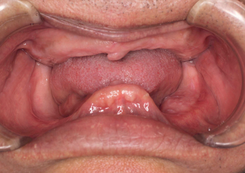
Fig 1: Starting situation: edentulous jaw. The posterior area of the lower jaw in particular showed advanced alveolar bone resorption.


ANATOMIC IM PRESSION AND PROVISIONAL RECORDING OF THE JAW RELATION
Following the evaluation of phonetics and function, the maxillomandibular relationship was provisionally recorded. Both the chin and nose were marked with a dot; then the distance between the dots was measured and the vertical dimension was established provisonally. In a next step, a double-arch impression was taken using Centric Tray (closedmouth impression). For this purpose, Centric Tray was loaded with silicone impression material that was given a slightly convex shape. After the tray had been seated in the mouth of the patient, we asked him to close the jaws at the “preliminary” vertical height and, after having performed the act of swallowing, to stay in this position until the impression material had set completely (Fig. 3).

As usual, the anatomical impression served as the basis for the fabrication of custom trays. In many cases, the preoperative situation is not sufficiently taken into account, as it is generally assumed that any deviations may be adjusted based on the functional impression later on. This can indeed be a misconception. If important areas are not covered by the functional tray, they are not captured in the impression. In order to select a suitable prefabricated impression tray, the jaw width is measured with a measuring loop. In the present case, the distance between the exterior of the maxillary tuberosities was measured in the upper jaw, and the distance between the mandibular tubercles in the lower jaw. A slightly overextended anatomical impression was made using a non-slumping impression material (Fig. 4).
FUNCTIONAL IM PRESSION, FINAL. BI TE REGI STRATION AND DETERMI NATION OF THE CENTRIC POSITION
Based on the existing information, custom trays were fabricated and handed over to the dentist. For the functional impression, a highviscosity silicone material (Virtual® Heavy Body) was used as the traymaterial, while a low-viscosity material (Virtual Light Body) was applied along the functional rim. The objective of the functional impression is to record the movements of the chewing muscles and facial muscles. This ensures that the denture borders precisely follow the contours of the oral mucosa. It should be kept in mind that denture “suction” is only achieved, if the borders of the denture adapt snugly to the mucous membrane (valve rim). In the present case, the suction effect was already noticeable when the impression was removed from the mouth after the impression material had set (Figs 5 and 6).
DETERMI NATION OF THE MAXILL OMANDIB ULAR RELATIONSHIP
When the BPS method is used, the joint related centric position of the mandible is determined by means of a horizontal maxillomandibular relationship record. However, a problem arises when the well-proven method of needle point tracing according to Gerber is applied in edentulous patients: How can the templates for the record be fixed in the mouth in a stable position? The Biofunctional Prosthetic System provides a solution to this problem: it comprises the intraoral registration


device Gnathometer M. Gnathometer M is prepared for needle point tracing based on a closed-mouth impression. This was also done in the present case. First, the device was correctly positioned in the mouth by means of a mounting jig. Then the needle point tracing record was made (Figs 7 and 8).
Suitable denture teeth were selected during the same appointment. We generally believe that it is very important to involve the patient in the selection process. Information about the patient’s satisfaction with previous tooth moulds, shades and tooth positions is also very helpful.In addition, a form selector was used (Phonares II FormSelector), which assisted in determining the suitable tooth moulds based on the patient’s interalar width. The Phonares denture tooth line allows teeth to be selected that are matched to the individual characteristics of the patient. Even though these teeth are “prefabricated”, they do not look like they are ready-made. Thus dentures result that look customized andare far from having an off-the-shelf appearance. We left it entirely to the patient to select the tooth shade and only offered advice (Fig. 9).
SET-UP AND COMPLETION OF THE DENTURES
In the set-up of the teeth, static and functional requirements as well asthe patient’s wishes were taken into account. The elderly man asked for an even front tooth row without any forced crowding. Due to tooth moulds matched to the age of the patient we were able to fulfil this request whilst avoiding an “unnatural” look. However, not only the align-ment of the teeth significantly influences the esthetic appearance. By creating a vestibular gingival portion that has a delicate yet effective appearance, the customized look can be accentuated with little extra
effort. In the present case, we used five different shades of wax to faithfully mimic the natural oral soft tissues (Fig. 10).
The wax-up was then tried in the mouth and evaluated on the basis of the following criteria: esthetics, phonetics, occlusal vertical dimension and centric relation. The patient was completely satisfied with the result and no modifications were required.
Following completion of the wax-up, we proceeded to process the denture base. The IvoBase denture base system allows wax dentures to be converted to resin in an easy and straightforward manner. The injection-based system ensures precise and reliable results which do not exhibit any inaccuracies. The models with the waxed denture bases were immersed in water, isolated and then flasked. One of the major advantages of the IvoBase system is that both the monomer and polymer are supplied in predosed capsules. Consequently, an accurate mixing ratio is ensured. Moreover, there is no risk of skin contact during mixing or danger of inhaling monomer vapours. For the case at hand, we chose the High Impact version (IvoBase, basic shade 34-V) which demonstrates high fracture toughness and is thus ideally suitable for removable dentures. After the flasks had been prepared (Fig. 11), they were placed in the injector and the program was started. The fully automatic injection and polymerization process is optimally adjusted to the properties of the IvoBase material; a continuous supply of material ensures that polymerization shrinkage is fully compensated.
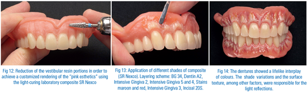
Subsequently, we devoted our attention to reconstructing the soft tissues. With the appropriate materials, the “pink esthetics” modelled in wax can be reliably converted to resin. The light-curing laboratory composite SR Nexco is perfectly suitable for this purpose. It ideally complements the IvoBase denture base material (shade 34-V) and offers dental lab technicians a wide array of design possibilities. After reducing the vestibular areas (Fig. 12), we applied different shades of SR Nexco. What is so remarkable about this composite is that it is available in a wide range of shades and offers outstanding sculptability (Fig. 13). We succeeded in mimicking the typical surface details of natural gingiva such as delicate stipplings, gentle alveolar bulges and slight recessions. After proper finishing and smoothing of the sharp edges as well as polishing, natural-looking dentures were achieved that demonstrated a lifelike interplay of colours. The different gingiva shades (SR Nexco) that had been used and the textured surface with its alternating convexities and flat areas were responsible for the light reflections along the denture base (Fig. 14). The teeth looked like they had actually “erupted” from the gingiva, just as in the natural, dentate jaw.
FINAL SEATING OF THE DENTURES
The fact that the patient showed a relaxed and satisfied smile whenthe dentures were seated for the first time was the first sign of success for the dental team (Fig. 15). After all the functional, esthetic and static criteria had been checked, we all agreed that the result was very appealing and would fulfil the patient’s needs for a long time. The dentures showed a stable intraoral fit. The patient was able to speak properly. Moreover, the assessment of the static and dynamic occlusion did not reveal any interferences, so that no adjustments had to be made. With much attention to detail, stateof- the-art materials and a proven setup protocol we were able to create dentures that matched the personality of the patient.

Fig 15: The patient with the dentures in place
CONCLUSION
Despite increased efforts to preserve natural teeth, the number of denture wearers throughout the world has remained constant. Due to the possibilities opened up by implant dentistry it is frequently assumed that the classical removable denture has lost significance. However, several objective factors contradict this assumption. Across the globe, the majority of edentulous patients are treated according to conventional methods. The BPS protocol, in combination with SR Phonares denture teeth, IvoBase denture base material and SR Nexco lab composite, is a reliable option that leads to predictable outcomes. The systematic, economical and stress-free procedure results in dentures that leave patients satisfied and help them regain their self-esteem and resume their social activities.
Disclaimer from Guident
The views and opinions expressed in articles published in Guident are solely those of the authors and do not necessarily reflect the views of the editors, the editorial board or the publisher, Ivory India. Guident does not endorse any product, technique or treatment described. Clinical content is intended for qualified dental and medical professionals and is not a substitute for individual clinical judgement. Guident, its editors and the publisher accept no liability for any consequence arising from the use of information published in the journal.
How to cite this article
Dr. Jiro Abe Clinical. FOR THE BENEFIT OF THE EDENTULOUS PATIENT, The Ideal Quartet For Complete Denture Fabrication: Methodology According To Bps, Sr Phonares Ii, Ivobase And Sr Nexco. Guident. 2013 Dec;7(1). Available from: https://ivoryindia.in/articles/prosthodontics/for-the-benefit-of-the-edentulous-patient-the-ideal-quartet-for-complete-denture-fabrication-methodology.html


