Non Surgical Management of Anterior Soft Tissue Defect
Deep Dental College, Faridabad (HAR) india
ABSTRACT
Periodontal surgical procedures may not always offer a predictable level of success in the reproduction of a natural gingival architecture. A different nonsurgical approach is described for the management of soft tissues deficiencies in anterior region. Among the various restorative treatments developed for the patient with an unaesthetic edentulous space is the fixed removable partial denture. Abutment tooth stabilization is combined with a removable partial denture to resolve challenging aesthetic problems.
KEYWORDS: Abutment, Fixed Partial Denture, Soft Tissue, Periodontal, Anterior
INTRODUCTION
The patient with a missing anterior teeth and a deficient alveolar ridge presents the problem of restoring the missing teeth and alveolar ridge. Numerous modalities of treatment are available to achieve the desired result. The choice depends on the complicating factors involved. A fixed partial denture may be the treatment of choice. In a situation with a soft tissue defect, however, a removable gingival facade might be used.1 The prosthesis should also be esthetic and allow the patient to maintain adequate oral hygiene. The various other treatment options available include Removable Partial denture with flange to mask the defect and Ridge augmentation with graft. Aesthetic surgical replacement is difficult and unpredictable, particularly when papillae in the aesthetic zone need to be restored.2 This article reports a conservative method for the replacement of anterior teeth associated with an anterior alveolar ridge defect. Porcelain fused to metal was used as the fixed partial denture segment and a modification of the Andrews type bridge was used for the removable segment. The prosthesis is designed to meet the requirements for aesthetics, comfort, phonetics, hygiene and favorable stress distribution to the abutments and soft tissue
CASE REPORT
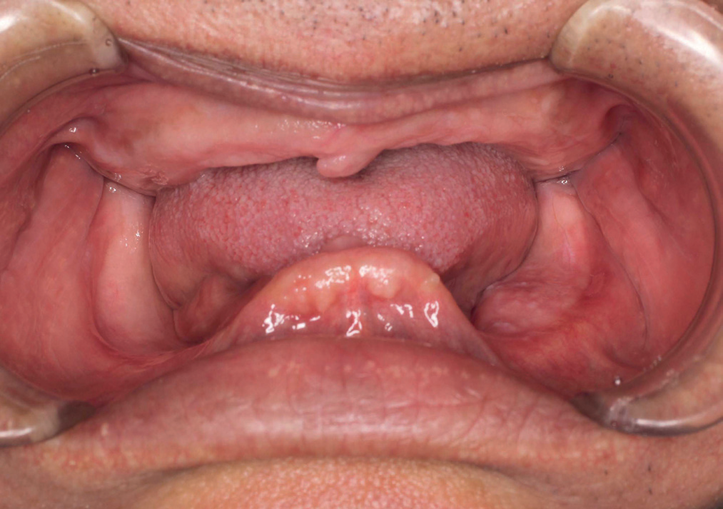
An 18 year old female reported to the Deep Dental Clinic, Faridabad, with a chief complaint of missing upper front teeth and wanted replacement. The patient met with an accident six years back, which led to the mobility of teeth and abscess formation in that region. Over the period of time there was bone resorption and loss of interdental papilla because of the chronic abscess. She got her mobile teeth extracted one year back (Fig 1a) and was wearing a removable partial denture since then. Teeth 11 & 12 were restored with custom-made post and core. During treatment planning, narrow width and reduced apical height of the residual alveolar ridge (i.e. Siebert’s Class III ridge relation) and a

Fig 1a
need to restore the teeth as well as lost supporting structures were taken into consideration. Keeping all these factors in mind, an Andrews Bridge was planned for the same.
TECHNIQUE
1. Patient’s diagnostic impression of the maxillary and mandibular arch was taken with irreversible hydrocolloid. Cast was poured in dental stone (Type III).
2. The appropriate bar was selected, from the preformed curvatures available using a diagnostic cast. The bar followed the residual ridge and was positioned in the approximate center of the replacement teeth.

3. Tooth preparations for porcelain fused to metal crowns were made on right central incisor and left canine having shoulder margin labially and chamfer margin lingually.
4. Increased reduction of the proximal surfaces adjacent to edentulous space was done to provide space for the mechanical lock of the bar in the crown and to reduce the possibility of distortion of the margins during soldering. (Fig. 1b)
5. After the abutments were prepared, gingival retraction was done. Final impression was made with a polyether impression material (Impregum, 3M ESPE). The impression was poured with Type V gypsum material (Die-Keen; Heraeus Kulzer, Armonk, NY). The working cast was prepared.
6. The wax patterns were contoured, and the bar positioned in relation to the crowns and the residual ridge. (Fig 2)
7. The completed wax patterns were invested. Casting was done followed by soldering. A sleeve was then placed over the bar, the teeth positioned and the flange completed in wax for the try-in. (Fig 3) The flange portion of the prosthesis was processed.
8. The esthetic try – in was done. The esthetics and tissue adaptation were evaluated. Followed by the cementation of the abutment retainers. The latitude in the placement of teeth and the opportunity for the try-in tend to eliminate the unaesthetic anti-smile line curve that occasionally occurs in fixed partial dentures because of technical requirements related to incisal relationships. (Fig 4)
9. The lingual surface of the finished restoration was completed. (Fig 5a, 5b & 6)
DISCUSSION
Patient selection is critical and problems that develop are essentially the result of diagnostic errors during treatment planning. A minimum of 2 mm vertical bar height is required for sufficient strength to support the removable portion of the restoration.3 Primary indications for this restoration are cases where the abutments are capable of supporting a fixed partial denture but the residual ridge has been partially lost due to trauma, congenital defects, or other pathologic process so that a conventional fixed partial denture would not adequately restore the patient’s missing teeth and supporting structures.
SUMMARY
Dentistry has many esthetic choices. A fixed removable bridge retained by an Andrew’s bar is an ideal choice for this situation. Conventional fixed bridges have many cosmetic, hygienic and functional disadvantages compared to Andrew’s bridges. Fixed pontic must be designed for cleaning in-situ.3,4 Tissue surfaces should be convex because concavities are impossible to clean and may ulcerate during flossing. Replacing papillae between pontic is an uncertain, drawn out surgical procedure. Unreplaced papillae feel un-natural and invite food impaction. Fixed bridges often result in very unaesthetic “black-holes”.4 Unlike Andrew’s bridges, all functional forces must be borne entirely by the abutments. The seating and closed adaptation of Andrew’s acrylic saddled on and around the ridge absorbs forces to abutments. Curved bars ensure that lifting or depressing forces are shared by all sectors. Plastic teeth supported in acrylic absorb shock and don’t wear the opposing dentition. Thus, this type of denture has qualities of both the fixed partial dentures and removable partial dentures and is indicated where the abutments would support a fixed partial denture but a severe defect is present in the edentulous space.
REFERENCES
- Mueninghoff A Leonard, Johnson H. Mark Fixed – removable partial denture. J Prosthet Dent 1982; 48(5) 547 – 50.
- Everhart J. Robert, Cavazos Edmund Evaluation of a fixed removable partial denture: Andrews Bridge System. J Prosthet Dent 1983; 50 (2) 180 – 84.
- Immekus E. Jameus and Aramany Mohamed. A fixed removable partial denture for cleft – palate patients. J Prosthet Dent 1995; 34(3) 286- 290.
- Andrews A. James, Biggs F. Walter the Andrews Bar and Sleeve Retained Bridge. A clinical report. Dentistry Today 1999;18(4).
Disclaimer from Guident
The views and opinions expressed in articles published in Guident are solely those of the authors and do not necessarily reflect the views of the editors, the editorial board or the publisher, Ivory India. Guident does not endorse any product, technique or treatment described. Clinical content is intended for qualified dental and medical professionals and is not a substitute for individual clinical judgement. Guident, its editors and the publisher accept no liability for any consequence arising from the use of information published in the journal.
How to cite this article
Dr. Vibha Kalra, Dr. Shikha Punjabi, Dr. Viram Upadhyaya. Non Surgical Management of Anterior Soft Tissue Defect. Guident. 2013 Dec;7(1). Available from: https://ivoryindia.in/articles/prosthodontics/non-surgical-management-of-anterior-soft-tissue-defect.html