
Prosthodontic Management of Edentulous Patients with Flabby Tissues: A Case Report
Department of Prosthodontics Post Graduate Institute of Dental Sciences, Pt. B.D Sharma University of Health Sciences, Rohtak, Haryana, India
ABSTRACT
A flabby ridge can be defined as a superficial area of mobile soft tissue affecting the maxillary or mandibular alveolar ridges. It can develop when hyperplastic soft tissue replaces the alveolar bone and is a common finding particularly in the maxillary anterior region of long term denture wearers. Masticatory forces may displace this mobile denture-bearing tissue, leading to denture instability and loss of peripheral seal. Forces exerted during impression making result in distortion of the mobile tissue. The appropriate management of flabby tissues by using special impression techniques is necessary to gain adequate support, retention and stability of complete dentures. This case report describes an impression technique for making impression of denture bearing areas containing flabby ridges.
Keywords: flabby tissue, impression technique, combination syndrome, Hobkirk technique
INTRODUCTION
Flabby ridge can be defined as a mobile soft tissue which is located on the superficial aspect of the maxillary and mandibular alveolar ridges.1 The reported prevalence has been demonstrated in upto 24% of edentulous maxillae and 5% edentulous mandibles.2 The objective of complete denture is to restore function, comfort and esthetics by replacement of missing teeth and alveolar structures using a stable prosthesis.
Displaceable flabby ridges, present a particular difficulty and may give rise to complaints of pain or looseness of the complete denture.3 Historically, flabby ridges found in the anterior maxilla were a feature of the ‘combination syndrome’.4,5 In this ‘condition’, the flabby tissue was thought to occur as a result of a maxillary complete denture opposing mandibular anterior natural teeth, without proper posterior occlusal support. Such flabby tissues could also arise due to unplanned or uncontrolled dental extractions.6
A master impression for a complete denture should record the entire functional denture-bearing area to ensure maximum support, retention and stability. Several impression techniques have been proposed in the literature for recording flabby ridges with the minimum amount of tissue displacement. The techniques for impression making in complete edentulous patients include, muco-compressive, muco-static and selective pressure impression technique.1 The mucostatic impression technique is used to record the mobile flabby tissues in undisplaced state.
CASE REPORT
A 65-year old male patient reported to the Department of Prosthodontics and Crown & Bridge with complaint of loose dentures. A thorough medical and dental history was taken. On examination, flabby tissue was found in the maxillary and mandibular anterior region extending from canine to canine region. Tissue blanching was also seen on pressure application [Figure 1a and 1b].

Figure 1a & 1b: Intraoral image of the maxillary and mandibular flabby ridge showing blanching
Fabrication of new set of complete denture was planned for the patient with recording of flabby tissue in undisplaced condition using Hobkirk technique.
The technique is as follows:
- The preliminary impression was made using irreversible hydrocolloid [Figure 2] (Zelgan, Dentsply) in perforated edentulous tray and the primary cast was poured in dental plaster. [Figure 3].
- Custom tray was fabricated using double spacer over the flabby tissue area. [Figure 4]
- After checking the proper tray extensions, border molding was done in conventional manner using green stick impression compound (DPI Pinnacle Tracing Sticks). [Figure 5]
- Spacer wax was removed and impression was made initially with zinc oxide eugenol impression paste (DPI impression paste). [Figure 6]
- The tray was then removed from the mouth and impression material was removed in the region of flabby tissue using a scalpel. Relief holes were made. [Figure 7]
- The tray was loaded in this region with light body poly vinyl siloxane material (Orikam, Neopure) and reseated in the patient’s mouth to record the flabby tissue. [Figure 8]
- Once set, the impression was removed from the mouth and inspected. Beading and boxing of the final impression was done using plaster pumice method and master cast was poured in Type III Dental stone. [Figure 9].
- Record bases were fabricated on the master cast followed by occlusal rim fabrication. Jaw relation was recorded in the usual manner.
- Following try-in, conventional complete denture was fabricated. [Figure 10]
- In subsequent visits, the patient reported with satisfactory retention, stability and function. [Figure 11]

Figure 2: Preliminary impression

Figure 3: Primary cast

Figure 4: Custom tray fabrication over double spacer

Figure 5: Border molding

Figure 6: Zinc oxide eugenol impression

Figure 7: Custom tray showing relief holes in the flabby tissue area

Figure 8: Final impression showing flabby tissue recorded in light body elastomeric impression material
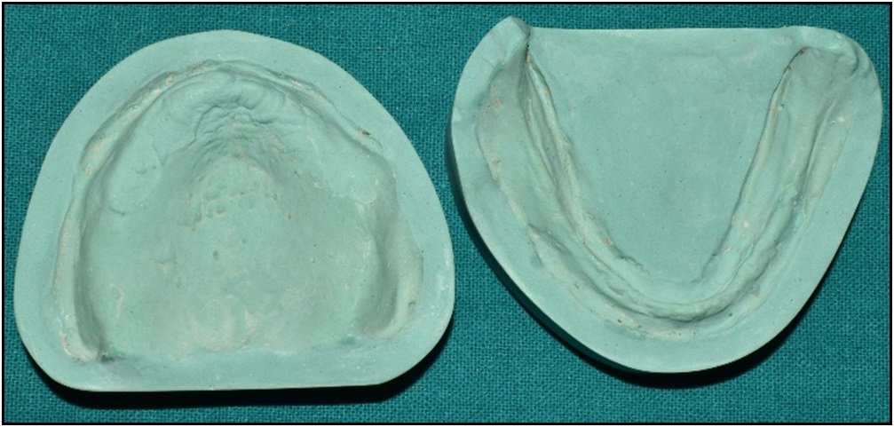
Figure 9: Master Cast

Figure 10: Intraoral image of Complete Denture

Figure 11: Pre-operative and Post-operative image
DISCUSSION
In 1972, Kelly and his colleagues described ‘combination syndrome’ caused by the presence of opposing natural teeth to an edentulous area. His observations included alveolar bone resorption in the anterior maxillary region, enlargement of the tuberosities and bone resorption underneath the mandibular denture bases.7
Multiple techniques for the management of flabby ridges have been described. Surgical methods include removal of flabby ridge using scalpel or by injecting a sclerosing agent prior to fabrication of complete denture. In addition, surgical ridge augmentation has also been proposed in the management of flabby ridges.1 As a result of accompanying medical conditions such elderly patients may not be suitable for surgically invasive procedures such as removal of flabby ridges, bone grafting, or placement of dental implants. The description of special impression technique is therefore timely. It describes how the management of poor denture-bearing areas can be accomplished by expanding on the basic principles of complete denture construction without using surgically invasive procedures.
In 1964, Liddlelow described a technique in which two separate impression materials were used in a custom tray (using ‘plaster of Paris’ over the flabby tissues and zinc oxide eugenol over the normal tissues). Osborne described a technique where two separate impression trays and materials were used to record the ‘flabby’ and ‘normal’ tissues separately and then related intra-orally. Watt and McGregor described a technique where impression compound was applied to a modified custom tray and a wash impression was made with zinc-oxide and eugenol.8-10
A common clinical challenge is the difficulty in uniform application and control of low viscosity impression materials on flabby tissues due to gravitational forces and different dental chair positions. Although Poly vinyl siloxane provides better accuracy for open window technique, controlled application is still a concern. Moreover, the modified window technique allows for controlled application of low viscosity impression materials in addition to the minimal exertion of pressure to the flabby ridges due to the presence of vents.1
CONCLUSION
Managing a patient with flabby ridge can be a challenging situation. Standard mucocompressive impression techniques are likely to result in an unretentive and unstable denture as the denture will be constructed on flabby tissues recorded in compressed state which will try to rebound and cause dislodgement of the denture. Mucostatic techniques may not make the best use of the available healthy tissue support. The use of selective pressure impression techniques helps to overcome some of these limitations by recording healthy tissues in compressed state thus gaining support from these tissues and recording flabby tissue in static form preventing denture instability during function.
REFERENCES
- Labban N. Management of the flabby ridge using a modified window technique and polyvinylsiloxane impression material. Saudi Dent J. 2018 Jan;30(1):89-93.
- Bansal R, Kumar M, Garg R, Saini R, Kaushala S. Prosthodontic rehabilitation of patient with flabby ridges with different impression techniques. Indian J Dent. 2014 Apr;5(2):110-3.
- Basker R M, Davenport J C. Prosthetic treatment of the edentulous patient. 4th edn. Oxford: Blackwell, 2002.
- Kelly E. Changes caused by a mandibular removable partial denture opposing a maxillary complete denture. J Prosthet Dent 1972; 27: 210-215.
- Lynch C D, Allen P F. The ‘combination syndrome’ revisited. Dent Update 2004; 31: 410-420.
- Lynch CD, Allen PF. Management of the flabby ridge: using contemporary materials to solve an old problem. Br Dent J. 2006 Mar 11;200(5):258-61.
- Kelly E. Changes caused by a mandibular removable partial denture opposing a maxillary complete denture. J Prosthet Dent 1972;27:140-50.
- Liddelow K P. The prosthetic treatment of the elderly. Br Dent J 1964; 117: 307-315.
- Osborne J. Two impression methods for mobile fibrous ridges. Br Dent J 1964; 117: 392-394.
- Watt D M, MacGregor A R. Designing complete dentures. 2nd edn. Bristol: IOP Publishing Ltd, 1986.
Disclaimer from Guident
The views and opinions expressed in articles published in Guident are solely those of the authors and do not necessarily reflect the views of the editors, the editorial board or the publisher, Ivory India. Guident does not endorse any product, technique or treatment described. Clinical content is intended for qualified dental and medical professionals and is not a substitute for individual clinical judgement. Guident, its editors and the publisher accept no liability for any consequence arising from the use of information published in the journal.
How to cite this article
Dr. Surbhi Mittal, Dr. Divya Dahiya, Dr. Sujata Chahal. Prosthodontic Management of Edentulous Patients with Flabby Tissues: A Case Report. Guident. 2023 Feb;16(3). Available from: https://ivoryindia.in/articles/prosthodontics/prosthodontic-management-of-edentulous-patients-with-flabby-tissues-a-case-report.html


