Prosthodontic Rehabilitation of a Patient with Hemimandibulectomy Using an Attachment Retained Partial Denture: A Clinical Report
Dharwad, Karnataka, India, Department Of Prosthodontics And Crown & Bridge, A.J. Institute of Dental Sciences, Mangalore. Karnataka. India
ABSTRACT
Rehabilitation of mandibular defects after resection following cancer surgery warrants multidisciplinary approach involving a maxillofacial prosthodontist, oral and maxillofacial surgeon, and a reconstructive surgeon. Occlusal disharmony, altered masticatory cycle, disfigured face, alteration of speech and low self-esteem are some of the challenges involved in rehabilitation of the hemimandibulectomy cases. The aim of this clinical report is to describe prosthetic rehabilitation of a patient following hemimandibulectomy using attachment retained cast partial denture to meet the functional and esthetic requirements.
Key words: Ameloblastoma, Hemi-mandibulectomy, Free Fibula Graft, Prosthetic rehabilitation, Metal framework, Mandibular prosthesis
INTRODUCTION
Ameloblastoma is considered as a most common tumor of odontogenic origin, which is a locally aggressive, slow-growing and persistent neoplasm of epithelial origin.1 Ameloblastoma is usually found in posterior mandible and surgical excision is the most common treatment.2 Surgical removal of a portion of mandible results in the gross alteration in the maxillomandibular relationship. There is resultant disfigurement of the face as residual mandibular fragment turns inward and backward in addition to tissue loss resulting from the removal of the tumor.3
Mandibular discontinuity defects are more debilitating than that portion of the mandible without loss of mandibular continuity.4 The reason for the deviation may be multifactorial and includes the remaining mandibular natural teeth, location and extent of the resection, the degree to which innervation has been involved, tightness of the wound closed by surgeon and the amount of soft tissue resected.5 The degree of impairment depends not only on the type of surgery and extent but also on the specific vulnerability of each function.3
Even though prosthodontic rehabilitation of patients with acquired mandibular defect is a challenging task for a dentist, prognosis for these patients has greatly improved with continued improvement in surgical resection and reconstruction techniques. Function, speech and appearance can be enhanced by prosthodontic management of patient with mandibulectomy defects.6 This article describes a technique of prosthodontic rehabilitation using attachment retained cast partial denture. The aim was to reposition the mandible in correct anatomical location and to enhance the patient’s confidence by improving the esthetics and function.
CLINICAL REPORT
A 49 year old female patient was referred to the Department of Prosthodontics for prosthetic rehabilitation after hemi-mandibulectomy following ameloblastoma. On clinical examination the mandible was resected from right sub condylar region to the body of the mandible on the left side and mandible was reconstructed using free fibula flap (Figure 1). Post-surgical panoramic radiograph (Figure 2) revealed reconstruction bone-plates from the right sub condylar region to left body of mandible.
Extra oral examination revealed deviation of mandible to the resected side leading to facial asymmetry. Intraoral examination revealed Kennedy Class II partially edentulous mandibular arch with remaining 17, 18, 19 and full complement of natural dentition in the maxillary arch. Resected right half of mandibular region showed thick, freely movable soft tissues with scar formation, loss of alveolar ridge and obliteration of buccal and lingual sulci. A treatment plan was formulated using attachment retained cast partial denture. Limitations of the prosthesis were clearly explained to the patient. Root canal treatment was carried out for 18, 19.

Fig 1: intraoral scar tissue

Fig 2: Panoramic radiograph
Diagnostic casts were obtained using irreversible hydrocolloid impression material (Jeltrate, Alginate, Fast set, Dentsply Philadelphia, PA, USA). Deflective occlusal contacts were corrected and composite (Multicore HB, Ivoclar/Vivadent AG, Liechtenstein) build up was done on the cuspal inclines of 14, 15, 18, and 19 so as to guide mandible into maximum intercuspation. Tentative centric jaw relation was recorded using wax occlusal rims. Putty index of lower arch was made to capture preoperative tooth anatomy with addition silicone material (Aquasil, Dentsply, USA). Tooth preparation was done on 18 and 19. Border moulding was done using heavy body vinyl polyvinyl siloxane impression material (Aqausil, Dentsply, USA) and final impressions were made using light body vinyl polysiloxane impression material (Aqausil, Dentsply, USA) to achieve maximum extension and tissue coverage (Figure 3). Provisional restorations were cemented using noneugenol cement (Provicol, Voco, Gemany). Impressions were poured with type IV die stone to get two sets of cast.
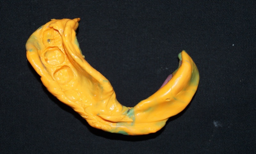
Fig 3: Final impression
Jaw relation was recorded using wax occlusal rims with temporary restoration in place to maintain vertical dimension. Phonetics and closest speaking space were used as a method for the determination of vertical dimension of occlusion. After establishing vertical dimension temporary crowns were removed and bite registration paste (Registrado X-TRA, Voco, Germany) was injected around the preparation to record the centric relation (Figure 4). Face bow transfer was done and casts were mounted on semi adjustable articulator (Hanau wide vue, Whip mix corporation). Wax patterns were fabricated on the die to prepare complete cast crowns. After completing the wax pattern, it was surveyed and the retentive arm of the clasp, rest seat, guiding plane and lingual bracing surface were prepared on the wax pattern (Figure 5).
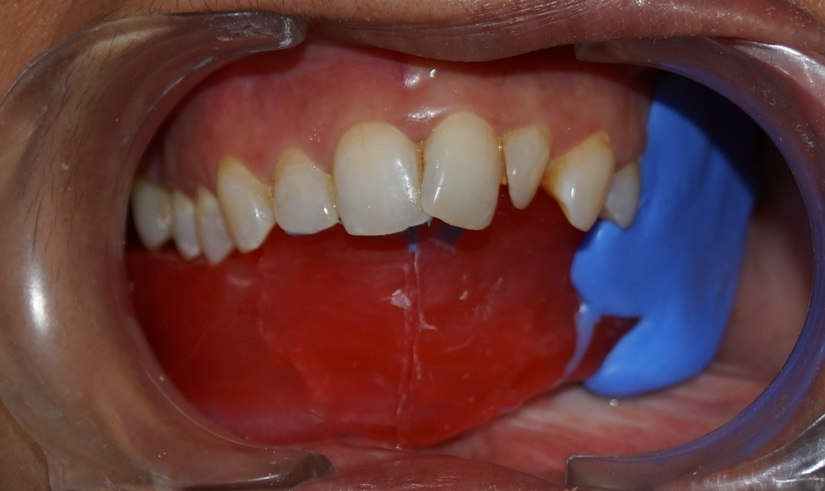
Fig 4: Bite registration
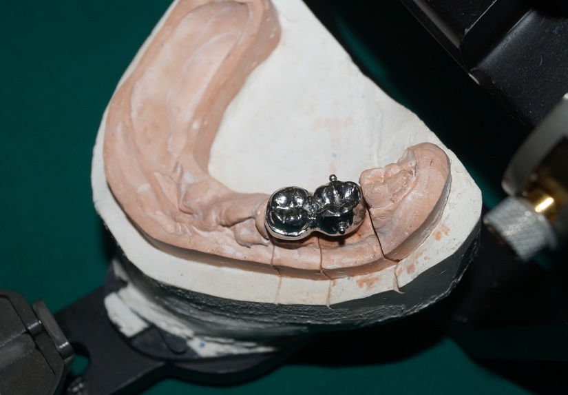
Fig 5 Rest seat and platform for the clasp arms on the crown
Placement of two Ceka Sagix attachment (Ceka) was planned on 18. Plastic1.7 mm spherical patrix was placed on center of buccal and lingual aspect of the wax pattern. The paths of the two patrixes were made parallel to each other. Wax patterns were cast and finished. The casting was surveyed again to ensure desired contours. A pink duplicating dummy were placed over the patrix and was aligned parallel. The undercuts were blocked. Wax up for the partial denture framework was done using LiWa Light-curing wax (LiWa; WP Bevern-Germany). Try in of the LiWa wax framework was done (Figure 6). Wax pattern was cast using lost wax technique without damaging the retentive ledge in the cavity for the matrix. Try in of the metal framework was done followed by the jaw relation.

Fig 6: Framework try in
As limited denture supporting area was available, it was planned to keep the teeth only upto the canine and balancing ramp was given on the defect side for cheek fullness and to prevent upper teeth from supraeruption. The trial dentures were evaluated for denture retention & stability during speech and eccentric jaw movements.
The dentures were processed in heat polymerizing acrylic resin as per manufacture’s instruction along with silicone based permanent soft lining material (Molloplast B, Detax GmbH, Ettlingen, Germany) in the defect area of the master cast. Removable partial dentures were finished and polished. Final insertion of the prosthesis (Figure 7) was done and patient was satisfied with the prosthesis.
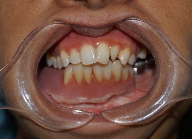
Fig 7: Attachment retained partial denture in-situ
Later, patient was trained to properly place and remove attachment retained cast partial denture and oral hygiene instructions were given to the patient. Recall visits were made at 1 week, 1 month, 3 month and 6 month interval.
DISCUSSION
Preci sagix is a sagittal ball attachment with segmented matrix mainly used for partial dentures and implant prosthesis. Advantages of sagix include cost effective, prolonged retention, patented snap mechanism, patient friendly and parts can be replaced easily. Also, unique keyway provides greater retention and stability due greater contacts area.7 Castable attachments with elastic retention are used to provide a more resilient and shock absorbing prostheses due to its elasticity.8 In this case abutment teeth were of inadequate clinical crown height to receive attachment; hence abutments were splinted posteriorly to edentulous span to aid in better distribution of stresses. Kapur et al have suggested by splinting teeth and full coverage crowns, adequate support can be achieved and it also improves overall prognosis of cast partial denture.9
Many techniques have been described in the literature to guide mandible to occlusion.10,11 In this case as the deviation was minor, occlusal interferences were removed and guide plane was adjusted until the splinted crowns occluded in maximum intercuspation.
Major connector was extended only till left premolar area where the underlining bone was healthy to make it more rigid and stable. 4 LiWa wax pattern was used for cast partial denture frame work, as it is dimensionally stable, accurate, avoids necessity of duplicate cast and allows wax framework trial.12
Only anterior teeth were arranged to enhance esthetics and balalcing ramp was given on the posterior side to prevent supraeruption of opposing teeth and patient discomfort on the resected site.13 Permanent soft denture liners due to their viscoelasticity and shock absorbing property offer effective solution for compromised and challenging edentulous situations.14,15,16 Hence, denture was lined with permanent soft denture liner on resected site in order to reduce the local stress concentration on the underlying grafted bone and tissues.
SUMMARY
Attachment retained cast partial dentures are a viable option for patients in whom implants and fixed prosthesis are contraindicated. Adherence to proper diagnosis and planning, precision techniques and periodic recall preventative therapy will result in long term success of treatment with preservation of the patient's existing dentition.
REFERENCES
- Gerzenshtein J, Zhang F, Caplan J, Anand V, Lineaweaver W: Immediate mandibular reconstruction with microsurgical fibula flap transfer following wide resection for ameloblastoma. J Craniofac Surg 2006; 17(1):178-82.
- Shafer WG, Hine MK, Levy BM, Tomich CE. A textbook of oral pathology, 4th ed. Philadelphia; WB Saunders; 1993. p. 86-229.
- Cantor R, Curtis TA .Prosthetic management of edentulous mandibulectomy patients -Part I Anatomic, physiologic and psycho logic considerations. J Prosthet dent 1971; 25:446-58.
- Taylor TD. Diagnostic considerations for prosthodontic rehabilitation of the mandibulectomy patient. In: Taylor TD, editor. Clinical maxillofacial prosthetics. Chicago; Quintessence Publishing; 2000. p. 155-70.
- Marunick M, Mahmassani O, Siddoway J, Klein B. Prospective analysis of masticatory function following lateral mandibulectomy. J Surg Oncol 1991; 47:92-7.
- Schneider RL, Taylor TD. Mandibular resection guidance prostheses: A literature review. J Prosthet Dent 1986; 55:84- 6.
- Waltz ML.Ceka extra coronal attachments. J Prosthet Dent 1973; 29(2):167-171.
- Feinberg E,Feinberg EM. Attachment retained partial denture. The New York State Dental Journal 1984; 161-64.
- Kapur KK, Deupree R, Robert J. Hasse AL.A randomized clinical trial of two basic removable partial denture designs. Part I: Comparisons of five-year success rates and periodontal health.J Prosthet dent 1994; 72(3):268- 82.
- Robinson JE, Rubright WC. Use of a guide plane for maintaining the residual fragment in partial or hemi-mandibulectomy. J Prosthet Dent 1964; 14:992-9.
- Sahin N, Hekimoglu C, Aslan Y. The fabrication of cast metal guidance flange prostheses for a patient with segmental mandibulectomy: A clinical report. J Prosthet Dent 2005; 93(3):217-20.
- Takaichi A,Wakabayashi N,Igarashi Y. Prefabricated light-polymerizing plastic pattern for partial denture framework. Contemp Clin Dent. 2011 Oct-Dec; 2(4):402-4.
- Desjardins RP,Occlusal considerations for the partial mandibulectomy patient. J Prosthet Dent 1979; 41(3):308-15.
- Bhargava A, Gupta S, Nagpal A, Kumar M, Chopra B.Innovative approach to bilateral maxillary undercuts using resilient soft liners - a case report.JIDA 2011; 12(5):1211-13.
- Wright PS. The success and failure of denture soft-lining materials in clinical use. J Dent 1984; 12:319-27.
- Bell DH. Clinical evaluation of a resilient denture liner. J Prosthet Dent 1970; 23:394-406.
Disclaimer from Guident
The views and opinions expressed in articles published in Guident are solely those of the authors and do not necessarily reflect the views of the editors, the editorial board or the publisher, Ivory India. Guident does not endorse any product, technique or treatment described. Clinical content is intended for qualified dental and medical professionals and is not a substitute for individual clinical judgement. Guident, its editors and the publisher accept no liability for any consequence arising from the use of information published in the journal.
How to cite this article
Dr. Mithun Upadhya, Dr. Satyabodh S. Guttal 2Sri Dharmasthala Manjunateshwara College of Dental Sciences & Hospital, Dr. Vidya K Shenoy. Prosthodontic Rehabilitation of a Patient with Hemimandibulectomy Using an Attachment Retained Partial Denture: A Clinical Report. Guident. 2022 Oct;15(11-12). Available from: https://ivoryindia.in/articles/prosthodontics/prosthodontic-rehabilitation-of-a-patient-with-hemimandibulectomy-using-an-attachment-retained-partial.html