LASER Disinfection and Retreatment of Failed Surgically Treated Anterior Teeth
Department of Conservative Dentistry Chandra Dental College and Hospital, Safedabad. Uttar Pradesh, India BBDCODS Lucknow, Uttar Pradesh
ABSTRACT:
The success of endodontic therapy is solidly based on efficient disinfection of the root canal system. Disinfection of root canal through use of laser radiation has been demonstrated by many studies including diode lasers. This article presents the case report of a 32 year old female who had undergone laser assisted non surgical management of failed root cannal filled teeth which had been twice treated surgically .
KEYWORDS: Root cannal, Diode Lasers, Laser disinfection
INTRODUCTION:
A conventional root canal treatment is always preferred over surgical endodontics and only when conventional root canal treatment fails we do surgical intervation. Endodontic retreatment offers the patient a second chance to save a root canal treated tooth that would otherwise be destined for extraction.
For the success of any root cannal therapy, the complete removal of the pathogenic bacterias and their toxic byproducts is of prime importance. Root canal bacteria, particularly Grampositive facultative anaerobes, seem to be remarkably resistant to local antimicrobial agents that are often used in root canal therapy. Nonthermal approaches to eliminate bacteria in endodontic treatments include sodium hypochlorite irrigation and photodynamic therapy. Routine sodium hypochlorite irrigation may remove the majority 98% of the bacteria within the canal but may not be very effective to bacteria located within the deeper layer of root dentin.1,2
Root canal bacteria, for example Enterococcus fecalis is a facultative anaerobic Gram positive coccus, which is present in oral flora is also related to the failure of endodontic treatment.3 Such bacterias seem to be remarkably resistant to any local antimicrobial agents that are often used in root canal therapy. Also , they form intra and extra radicular biofilms, which are difficult to remove and they cause persistent root canal infections or reinfection.4,5
Nonthermal approaches to eliminate bacteria in endodontic treatments include sodium hypochlorite irrigation and photodynamic therapy. Routine sodium hypochlorite irrigation may remove the majority 98% of the bacterias within the canal but may not be very effective to bacteria located within the deeper layer of root dentin.6,7
Lasers have been used in dentistry for removing hard tooth tissue for more than 20 years. Recently, laser systems have gained importance in the field of laser assisted endodontics, namely the Nd:YAG, the diode, the Er:YAG, and the Er,Cr:YSGG laser. They have also been proposed for use in root canal therapy for cleaning and for eliminating bacteria from the root canal system . The heat induced cytotoxic response kills bacteria in the root dentin via heat conduction from the laser incident root canal surface.8,9
The present case report describes the laser assisted non surgical management of failed root canal filled teeth which had previously been twice surgically treated.
CASE REPORT
A 32 yrs old female patient reported to the Department of Conservative and Endodontics in BBDCODS, Lucknow with the chief complaint of severe pain & swelling in lower anterior teeth since
3 days. She gave history of root canal treatment followed by root surgery in the same teeth 5 years back. Due to continuous discomfort and pus discharge, another endodontic surgery was performed 2 years back in the same teeth region.
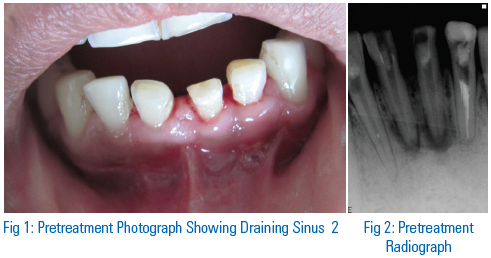
On intra-oral examination there was a draining sinus (Figure 1) in relation to 31 and 41 .The teeth were tender on purcussion with grade II mobility.
On radiographic examination, a well defined periapical radiolucency with insufficiently obturated canals were seen in relation to 31 and 41. The root apex were blunted suggestive of previous apicectomy but there were no evidence of any retrograde filling (Figure 2) .
The patient was explained about the existing condition and the need for retreatment was stressed upon. Patient agreed for orthograde retreatment but was skeptic about the surgical intervention. Therefore, it was decided to go for non surgical endodontic retreatment. Patient was explained about regular followups that were necessary for the evaluation of treatment.
TREATMENT PLAN
The old gutta percha were removed with the help of H-file and gutta percha solvent (Figure 3). The canals were thoroughly rinsed with normal saline and 2% chlorohexidine. Chlorhexidine was used as an irrigants it has demonstrated its broad spectrum antibacterial activity. After gutta percha removal we placed calcium hydroxide based intracanal medicament and temporized with zinc oxide


eugenol cement. Adjuvent to conventional therapy, Low Level Laser Therapy (LLLT) was delievered using diode laser at 0.8 watt in non contact mode in the lower anterior teeth region (Figure 4 ). The patient was recalled after one week.
Patient came back after 1 week with no pain, swelling and a healing sinus. Cleaning and shaping was performed by conventional method. Again intracanal medicament was placed followed by LLLT and patient was recall after 5 days for obturation.
After 5 days the patient was re-examined and the canals were dry and teeth were asymptomatic. At this point of time laser assisted disinfection was performed in each tooth using 810nm diode laser. The fiber tip with an outer diameter of 300 µm was inserted into the root canals at a distance of 1 mm from the apical foramen. The laser was operated in pulsed mode (15 Hz) at 1.5 watt output power in contact mode under constant scanning movement of the optical fibre at an angle of 10 degrees. One lasing cycle comprised five irradiations of 5 seconds each, with 15 seconds intervals without any water spray or air cooling (Figure 5).
Gutta percha were customised to snugly fit at the apex and the cannals were obturated using resin sealer to achieve better seal. The other option would have been to seal the apical part with MTA followed by obturation with gutta percha but this was not carried out due to economic constrain of the patient (Figures 5-8).

Patient was constantly reviewed if futher surgical intervention was necessary but it was observed that size of lesion gradually decreased suggesting healing of the lesion and there was no recurrence of symptoms when followed up for a period of 1 week , 1 month, 6 months and 12 months (Figure 9 (A-D) respectively).
DISCUSSION
A decision whether to retreat, restore or extract a failed root cannal filled tooth requires thorough examination and evaluation of the tooth. The reason many teeth do not respond to root canal treatment is because of procedural errors that prevent the control and prevention of intra canal endodontic infection.
Undoubtedly, the major factors associated with endodontic failure are the persistence of microbial infection in the root canal system and/or the periradicular area.10-13
Kouchi et al. 14 could demonstrate that bacteria are capable of invading the periluminal dentin up to a depth of 1,100 mm. On the other hand, Berutti et al15 explained that the chemical disinfectants penetrate no more than 130 mm into the dentin . The discrepancy of the penetration depth of micro-organisms and bactericidal rinsing solutions often holds responsible for therapy resistant cases and long-term failures which can be observed in conventional endodontics.
When the choice of non surgical endodontic retreatment is made then the primary goal is to access the pulp chamber and remove any microbial infection in the root canal system and/or the periradicular area or repair any defects that are pathological or iatrogenic in origin.
The introduction of lasers in endodontics has dramatically improved the effectiveness and success rate of root canal treatment. Dental lasers provide greater accessibility of formerly unreachable parts of the tubular network due to their better penetration into dentinal tissues.16,17
Generally, in any case surgical retreatment would be carried out following a nonsurgical endodontic failure. But in this case, patient had already undergone surgical intervention twice and totally refused to experience another traumatic experience.
Hence it was decided to perform a nonsurgical retreatment along with thorough disinfection with diode laser and effective sealing of infected root canal.
Scientific research was first conducted with the Nd:YAG and the diode lasers which gained widespread acceptance in the fields of laser-assisted endodontics.18-23
In recent years numerous authors have conducted various studies on different laser systems in endodontic treatment and found lasers as an effective tools for root canal cleansing and disinfection. Moritz et al. 20 determined that irradiation with a Diode laser in two subsequent sessions resulted in almost complete elimination of bacteria. Schoop et al.22 reported that Nd:YAG and Diode lasers at 1W are efficacious for E. faecalis .
Also, it has been observed that the non surgical retreatment shows improved outcomes with increasing recall time.24,25
CONCLUSION
There is enough potential for success of primary root canal filling but fact remains that clinicians are confronted with post treatment disease. Diode laser has been tested with promising results in relation to dentinal disinfection. Because of lasers properties and its low cost in relation to most lasers used in dentistry it has proved to be a resource worth testing. Further clinical studies should be encouraged to assess the possible improvements of endodontic treatment using Nd:YAG and Diode laser radiation.
REFERENCES
- Kouchi Y, Ninomiya J, Yasuda H, Fukui K, Moriyama T, Okamoto H. Location of streptococcus mutans in the dentinal tubules of open infected root canals. J Dent Res. 1980;59(12): 2038–2046.
- Berutti E, Marini R, Angeretti A. Penetration ability of different irrigants into dentinal tubules. J Endod. 1997; 23(12):725–727.
- Radatti DA, Baumgartner JC, Marshall JG. A Comparison of the Efficacy of Er,Cr:YSGG Laser and Rotary Instrumentation in Root Canal Debridement. J. Am. Dent. Assoc. 2006;137 (9): 1261–1266.
- Gordon W, Atabakhsh VA , Meza F , Doms A, Nissan R, Rizoiu I , Stevens RH. The Antimicrobial Efficacy of the Erbium, Chromium: Yttrium-Scandium-Gallium-Garnet Laser With Radial Emitting Tips on Root Canal Dentin Walls Infected With Enterococcus Faecalis. J. Am. Dent. Assoc. 2007;138(7): 992–1002.
- Siqueira JF Jr, Lopes HP. Bacteria on the apical root surfaces of untreated teeth with periradicular lesions: a scanning electron microscopy study. International Endodontic Journal 2001; 34: 216–220.
- Sundqvist G, Fidgor D, Persson S, et al. Microbiologic analysis of teeth with failed endodontic treatment and the outcome of conservative retreatment. Oral Surg Oral Med Oral Pathol Oral Radiol Endod 1998;85: 86–93.
- Nair PNR, Sjögren U, Krey G, Kahnberg KE, Sundqvist G. Intraradicular bacteria and fungi in root-filled, asymptomatic human teeth with therapy-resistant periapical lesions: a long-term light and electron microscopic follow-up study. Journal of Endodontics 1990a ;16: 580–8.
- Lin LM, Skribner JE, Gaengler P. Factors associated with endodontic treatment failures. Journal of Endodontics 1992; 18,:625–7.
- .Kouchi Y, Ninomiya J, Yasuda H, Fukui K, Moriyama T, Okamoto H. Location of streptococcus mutans in the dentinal tubules of open infected root canals. J Dent Res 1980;59(12): 2038–2046.
- .Berutti E, Marini R, Angeretti A. Penetration ability of different irrigants into dentinal tubules. J Endod 1997; 23(12):725–727.
- .Klinke T, Klimm W, Gutknecht N. Antibacterial effects of Nd:YAG laser irradiation within root canal dentine. J Clin Laser Med Surg 1997;15:29–31.
- Odor TM, Chandler NP, Watson TF, Ford TR, McDonald F. Laser light transmission in teeth: A study of the patterns in different species. Int Endod J 1999;32(4):296–302.
- 13. Myers TD, McDaniel JD. The pulsed Nd:YAG laser: Review of clinical applications. J Calif Dent Assoc 1991; 19(11):25–30.
- Moritz A, Gutknecht N, Goharkhay K, Schoop U, Wernisch J, Sperr W. In vitro irradiation of infected root canals with a diode laser: Results of microbiologic, infrared spectrometric and stain penetration examinations. Quintessence Int 1997; 28:205–209.
- Moritz A, Gutknecht N, Schoop U, Goharkhay K, Doertbudak O, Sperr W. Irradiation of infected root canals with a diode laser in vivo: Results of microbiological examinations. Lasers Surg Med 1997;21:221–226.
- Gutknecht N, van Gogswaardt D, Conrads G, Apel C, Schubert C, Lampert F. Diode laser radiation and its bactericidal effect in root canal wall dentin. J Clin Laser Med Surg 2000;18(2):57–60.
- Schoop U, Kluger W, Moritz A , Nedjelik N, Georgopoulos A , Sperr W. Lasers Surg. Med. 2004; 35:111-116.
- Hardee MW, Miserendino L, Kos W, Walia H. Evaluation of the antibacterial effects of intracanal Nd:YAG laser irradiation. J Endod. 1994;20(8):377–380.
- Cristian et al.Treatment outcome in endodontics: The Toronto study-phases 3 and 4: Orthograde retreatment. J Endod 2008;34:131-137.
- Torabinejad et al. Outcomes of non surgical retreatment and endodontic surgery: A systematic review. J Endod 2009;35:930-7.
Disclaimer from Guident
The views and opinions expressed in articles published in Guident are solely those of the authors and do not necessarily reflect the views of the editors, the editorial board or the publisher, Ivory India. Guident does not endorse any product, technique or treatment described. Clinical content is intended for qualified dental and medical professionals and is not a substitute for individual clinical judgement. Guident, its editors and the publisher accept no liability for any consequence arising from the use of information published in the journal.
How to cite this article
Dr. V. K. Srivastava, Dr. Shally Mahajan, Dr. Akanksha Bhatt. LASER Disinfection and Retreatment of Failed Surgically Treated Anterior Teeth. Guident. 2013 Nov;6(13). Available from: https://ivoryindia.in/articles/endodontics/laser-disinfection-and-retreatment-of-failed-surgically-treated-anterior-teeth.html