Non -Sur Gical Endodontic Treatment of Periapical Lesions Using Calcium Hydroxide
Department of Conservative Dentistry And Endodontics Sharad Pawar Dental College Datta Meghe Institue Of Medical Sciences (Du) Wardha, India
ABSTRACT
The success of root canal treatment is based on total elimination of root canal content, thorough cleaning, shaping and obturation of root canal system. For the success of endodontic treatment, healing of periapical pathology is necessary and can be achieved by maintaining a sterile root canal system. Calcium hydroxide has the potential to maintain a sterile root canal by its antibacterial and tissue-dissolving property. In these case reports, root canal treatment proved successful in promoting the healing of periapical lesions. This confirms that periapical lesions can respond favorably to non-surgical treatment.
KEY WORDS: calcium hydroxide, nonsurgical endodontic treatment, periapical healing, periapical lesion.
INTRODUCTION
Bacterial infection of the dental pulp may lead to periapical lesions. They are generally diagnosed either during dental radiographic examination or following acute pain in tooth.Most periapical lesions can be classified as dental granulomas, radicular cysts or abscesses. The incidence of cysts within periapical lesions varies between 6 and 55%[1]. The occurrence of periapical granulomas ranges between 9.3 % and 87.1%, and of abscesses between 28.7% and 70.07%. There is clinical evidence that as the periapical lesions increase in size, the proportion of the radicular cysts increases. Periapical lesions are a sequelae to endodontic infection caused due to dental caries or trauma and manifests itself as the host defense response to microbial challenge emanating from the root canal system. It is viewed as a dynamic encounter between microbial factors and host defenses at the interface between infected radicular pulp and periodontal ligament that results in local inflammation, resorption of hard tissues, destruction of other periapical tissues.[2]
The ultimate goal of endodontic therapy should be to return the involved teeth to a state of health and function without surgical intervention.All inflammatory periapical lesions should be initially treated with conservative nonsurgical procedures[3]. Surgical intervention is recommended only after nonsurgical techniques have failed . A high percentage of 94.4% of complete and partial healing of periapical lesions following nonsurgical endodontic therapy has also been reported. Various chemicals such as phenols, aldehydes, antibiotics, iodine, and calcium hydroxide have been used as intracanal medicaments. No agents have yet satisfied all the requirements of the medicament as outlined by Grossman et al, particularly in relation to the irritation of the periradicular tissue, systemic distribution, and allergic reaction.Calcium hydroxide is a material that has been used for a variety of purposes since its introduction into dentistry. In its pure form, calcium hydroxide has a high pH, and its dental use relates chiefly to its ability to stimulate mineralization and also to its antibacterial properties.[4] Recent studies have recommended calcium hydroxide as a root canal medicament because of its low toxicity, bactericidal activity, and biocompatibility. Few reports have discussed the tissue-dissolving effect of calcium hydroxide. Hence, calcium hydroxide has been suggested as a root canal medicament that would enhance the healing of periradicular tissue.
CASE 1
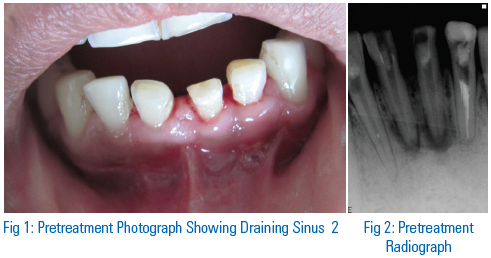
A 50-year-old female reported to the department of conservative dentistry and endodontics with a chief complaint of localized swelling and pus discharge in the lower left back region of the jaw since 3weeks. Clinical examination revealed localized swelling in relation with mandibular left first molar (tooth-36) with presence of sinus tract and discharge of pus (Fig.A,B). Electric and thermal pulp testing showed no response with 36.Endodontic treatment was planned with 36. Non surgical endodontic treatment was proposed with patient’s consent and patient was explained about the treatment and it’s outcome. On first visit access cavity was prepared on 36.Working length was determined and canals were cleaned and shaped by Kerr-files (MANI, Inc Japan) using step back technique.(Fig. C)


Irrigation was done using 3% sodium hypochlorite (NOVO Dental Pvt. Ltd., India). Intracanal medicament of calcium hydroxide powder mixed with chlorhexidine was placed in canal using lentulo spiral. Access cavity was closed with interim dressing. Patient was kept on follow up and intracanal calcium hydroxide dressing was changed periodically. Significant reduction in size of periapical lesions was seen at periodic follow up visits. The tooth was symptomless and so root canal was obturated with gutta-percha points and a calcium hydroxide-based root canal sealer (Sealapex; Sybron/Kerr, Romulus, MI, USA) using the lateral condensation technique .(Fig-D) .
The patient remained symptomless during the recall appointments at 3 months,6 months and 9 months.(Fig E-H)
CASE 2:
A 42 years old male patient reported to the department of conservative dentistry and endodontics with the chief complaint of pain and mobility in lower front teeth since 2 months. Past history revealed trauma 9 years back. On clinical examination discoloration was seen in relation to 31, and grade-I mobility in relation to 31(Fig-I). Electric and thermal pulp testing showed no response in 31. Intraoral Periapical Radiograph showed bone loss distal aspect and ill-defined radiolucency around the apices of 31 suggestive of Chronic periapical pathology in 31.Provisional diagnosis was made as Chronic Periapical abscess in relation to 31. Hence non surgical endodontic treatment was proposed with patient’s consent. On

first visit access cavity was prepared on 31.(Fig-J).Working length was determined and canals were cleaned and shaped by Kerr-files (MANI, Inc Japan) using step back technique.
Irrigation was done using 3% sodium hypochlorite (NOVO Dental Pvt. Ltd., India). Intracanal medicament of calcium hydroxide powder mixed with chlorhexidine was placed in canal using lentulo spiral. Access cavity was closed with interim dressing. Patient was kept on follow up and intracanal calcium hydroxide dressing was changed periodically(Fig-J). Significant reduction in size of periapical lesions was seen at periodic follow up visits. The tooth was symptomless and so root canal was obturated with gutta-percha points and a calcium hydroxide-based root canal sealer (Sealapex; Sybron/Kerr, Romulus, MI, USA) using the lateral condensation technique .(Fig-K).
The patient remained symptomless during the recall appointments at 3 months,6 months and 9 months.(Fig-L-N)
CASE 3
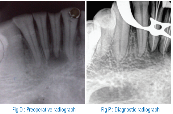
A 38 years old male patient reported to the department of conservative dentistry and endodontics with the chief complaint of pain and intermittent pus discharge in lower front tooth region since 10 days. On clinical examination sinus tract was present in relation to 31, 41, and the teeth were tender on percussion (Fig.O). On electric and thermal pulp testing was positive with 31 and 41 . Intraoral Periapical Radiograph revealed periapical radioluceny in 31, 41, region. Provisional diagnosis was made as chronic periapical lesion in relation to 31,and 41. Endodontic treatment was planned and executed in 31, 41 with informed patient’s consent. On first visit access cavity was prepared on 31and 41. Working length was determined and two canals were located with 31 and 41.(Fig-P)The canals were cleaned and shaped by Kerr-files (MANI, Inc Japan) using step back technique. Irrigation was done using 3% sodium hypochlorite (NOVO Dental Pvt. Ltd., India) and metrogyl. Intracanal medicament of calcium hydroxide powder mixed with chlorhexidine was placed in canal using lentulo spiral. (Fig-Q,R).Access cavity was closed with interim dressing. Patient was kept on follow up and intracanal calcium hydroxide dressing was changed periodically. Significant reduction in size of periapical lesions was seen at periodic follow up visits. The tooth was symptomless and so root canal was obturated with gutta-percha points and a calcium hydroxide-based root canal sealer (Sealapex; Sybron/Kerr, Romulus, MI, USA) using the lateral condensation technique (Fig-S,T).
The patient remained symptomless during the recall appointments at 6 and 9 months.(Fig-U,V)


DISCUSSION
A nonsurgical approach should always be adopted before resorting to surgery. When there is no drainage of fluid from the canals, calcium hydroxide or the triple antibiotic paste can prove beneficial. Periodic follow-up examinations can be used to monitor the healing of periapical lesions[5] .
Periapical lesions of endodontic origin may develop asymptomatically and become large. Proper biomechanical preparation followed by calcium hydroxide medication renewed periodically represents a nonsurgical approach to resolve extensive inflammatory
periapical lesions.
The conservative treatment success in managing supposedly cystic
periapical lesions could be explained based on the following aspects:
- Biomechanical preparation and bacterial control
- Lesion decompression achieved by apical patency
- Complementary antiseptic action of calcium hydroxide due to its alkalinity and its bony repair effect.
- Good immune response due to patients age[6].
The exact mechanism by which periapical cysts heal is also not clearly understood. As the lumen of a ‘bay’ or ‘pocket’ cyst is open to the root canal, it is likely to heal after conventional root canal treatment due to the removal of intra-canal irritants. In contrast, the tissue dynamics of a true cyst are self-sustaining by virtue of its independence of the presence or absence of irritants in the root canal.
True cysts, particularly large ones, containing cholesterol crystals are less likely to be resolved by conventional root canal treatment.
Because it is clinically and radiographically impossible to differentiate a bay cyst from a true cyst, as it is likewise between a cyst and a granuloma, judicious treatment planning should favour a conservative approach to treatment[7] .
A long-standing endodontic infection allows bacteria to propagate throughout the root canal system, which plays an essential role in pulpoperiradicular pathosis. Biochemical cleaning and shaping of the root canal with the assistance of antimicrobial irrigants may fail to eliminate bacteria from the root canal system. Residual bacteria and their by-products negatively affect the repair of the periradicular apparatus.[40] The use of biocompatible intracanal medication possessing antimicrobial properties may reduce or eliminate bacteria in the root canal system and significantly increase the success of root canal therapy.
Caliskan and Turkum reported periapical repair and apical closure of a pulpless tooth using calcium hydroxide powder mixed with saline. Oztan[8] used calcium hydroxide powder mixed with saline as an intracanal medicament in which large periapical lesions were completely
healed within a period of 3 months. Donald also used saline to mix calcium hydroxide
powder as an intracanal medicament. Souza et al[9] advocated that direct contact between calcium hydroxide and the periapical tissue was necessary for an adequate inductive action. Souza et al also suggested that the action of calcium hydroxide beyond the apex may be 4-fold: (1) anti-inflammatory through its hygroscopic action, (2) neutralization of acid products such as hydrophase, (3) activation of alkaline phosphatase, and (4) antibacterial action. Caliskan and Sen suggested that teeth with periapical lesions in which calcium hydroxide paste was extruded did not show a different healing pattern from the one treated conventionally.Weiger cited the influence of calcium hydroxide intracanal dressing on the progress of teeth with endodontically induced periapical lesions. In the present case report, calcium hydroxide powder was mixed with saline to form a paste that was used as intracanal medicament in accordance with Caliskan and Oztan. Heithersay recommended the use of intracanal calcium hydroxide dressing in teeth with a large periapical lesion. Hasselgren stated that calcium hydroxide applied to dissolved necrotic material from the periapical region and root canals ultimately helps in healing of periapical tissue.
Bystrom et al [10] reported that calcium hydroxide has the potential to kill bacteria left in an inaccessible area of the root canal system and therefore favors healing of periapical tissue. Lynne suggested that 10% calcium hydroxide may be more effective than Peridex or 10% calcium hydroxide in Peridex for theelimination of E faecalis from dentin tubule,
which has been found to be present in 38% ofteeth with endodontic failure.
Fisher and McCabe stated that the bactericidal property of calcium hydroxide is directly proportionalto the ability of calcium hydroxide to diffuse from set material.
CONCLUSION
Root canal treatment with calcium hydroxide as intracanal medicament
proved successful in promoting the healing of a periapical lesion. But a long follow-up is necessary to access the healing of the defects. Thus non surgical treatment will be the preferred choice because it seems to provide the most benefit with the lowest risk.
REFERENCES
- Nair PNR, Pajarola G, Schroeder HE. Types and incidence of human periapical lesions obtained with extracted teeth. Oral Surg Oral Med Oral Pathol Oral Radiol Endod 1996; 81:93-102.
- Nair P.N.R. Apical periodontitis: a dynamic encounter between root canal infection and host response. Periodontology 2000, 1997; 13(1) : 121-148
- Lin LM, Huang GT, Rosenberg PA. Proliferation of epithelial cell rests, formation of apical cysts, and regression of apical cysts after periapical wound healing. J Endod 2007;33:908-16.
- Foreman PC, Barnes JE.A review of calcium hydroxide. Int Endod 1990;23:283–297.
- Nair PNR, Pajarola G, Schroeder HE. Types and incidence of human periapical lesions obtained with extracted teeth. Oral Surg Oral Med Oral Pathol Oral Radiol Endod 1996; 81:93-102
- García CC, Sempere FV, Diago MP, Bowen EM.The post-endodontic periapical lesion: histologic and etiopathogenic aspects. Med Oral Patol Oral Cir Bucal. 2007;12(8):E585-90.
- 7. Caliskan MK. Prognosis of large cyst-like periapical lesions following nonsurgical root canal treatment. Int Endod J 2004; 37: 408–16.
- Bystrom A, Claesson R, Sundqvist G.The antibacterial effect of camphorated paramonochlorophenol, camphorated phenol and calcium hydroxide in the treatment of infected root canals. Endod Dent Traumatol 1985;1:170–175
- Oztan MD. Endodontic treatment of teeth associated with a large periapical lesion. Int Endod J 2002;35:73–78
- Souza V, Bernabe PFE, Holland R, Nery MJ,Mello W, Otobonl Filho JA.Tratamento nao cururgico de dintis com lesos periapicais. Rev Brasil Odontol 1989;46:39–46
Disclaimer from Guident
The views and opinions expressed in articles published in Guident are solely those of the authors and do not necessarily reflect the views of the editors, the editorial board or the publisher, Ivory India. Guident does not endorse any product, technique or treatment described. Clinical content is intended for qualified dental and medical professionals and is not a substitute for individual clinical judgement. Guident, its editors and the publisher accept no liability for any consequence arising from the use of information published in the journal.
How to cite this article
Dr. Keyur P. Chande, Dr. Narendra U. Manwar, Dr. Manoj G. Chandak. Non -Sur Gical Endodontic Treatment of Periapical Lesions Using Calcium Hydroxide. Guident. 2013 Nov;6(13). Available from: https://ivoryindia.in/articles/endodontics/non-sur-gical-endodontic-treatment-of-periapical-lesions-using-calcium-hydroxide.html