Prosthodontic Rehabilitation of Malpositioned Implants After Ameloblastoma Followed by Mandibulectomy and Fibula Bone Graft
Consultant Implantologist, India
ABSTRACT
The clinical report describes the rehabilitation with fibula bone graft reconstruction and 2 implants of a patient with a partial mandibular defect as a result of a partial mandibulectomy due to ameloblastoma of the left mandible. Due to the altered shape of the graft bone, the implants were malpositioned in the buccolingual plane.Using an implant-supported screw retained hybrid acrylic prosthesis we successfully completed the prosthodontics rehabilitation. This implant-supported prosthesis successfully restored function and esthetics. Aspects that deal with the repair of the buccal soft tissue are discussed.
Key words: fibula bone graft, malposition, and implant supported prosthesis.
INTRODUCTION
Defects of the mandible may result from treatment of benign and malignant tumours, congenital abnormalities, trauma, infections, and osteoradionecrosis. The ideal goal of reconstruction is to achieve sufficient anatomical bulk (height and width) of the mandible that will accommodate implant insertion and adequate muscle attachments to allow for normal form and function, preservation of the inferior alveolar nerve and subsequent restoration of sensation.1 Restoration of the form (esthetics) and function (mastication, deglutition, and saliva control) have been achieved by the use of autogenous grafts harvested from the iliac crest, rib, radius, fibula, tibia, scapula, and calvarium.1
Vascularized bone flaps, in general, provide the best functional and aesthetic outcome, with the fibula flap remaining the gold standard for mandible reconstruction.2
Studies show implant-support- edoverdentures have superior retention to conventional dentures. Regardless of the type of attach- ment system used -- bar, ball or magnet -- patients are significantly more satisfied with implant-sup- ported overdentures than with con- ventional dentures.3
The benefits of implant-retained prosthesis have been recognized since several years. Dental implants may improve denture retention and stability without unnecessaryloading of the vulnerable mucosa. Function, comfort, aesthetics, and eventually the quality of life can be improved.4,5
They can restore severe alveolar defects and replace lost dentition and are removable for hygienic purposes. 6 Ball & bar attachments are the most commonly used retainer system used for implant-bearing overdentures.7 They act as a rigid anchorage & provide the overdentures with advantage that are offered by both removable and fixed prostheses. Thus, implant supported prostheses may ensure that the patient can accomplish significantly better masticatory performance on the side of the mouth that has the defect.
However, osseointegration of implants into the graft bone represents only the primary, but not the only, requirement for a successful implant-supported prostheses.8 It is still a challenge for prosthodontists because of insufficient bone height and poor quality overlying soft tissue. This clinical report describes the prosthodontics rehabilitation of a patient with partial mandibular defect and malpositioned implants after a fibula bone graft and describes a new way to rehabilitate patients who find themselves in such unsatisfactory situations.
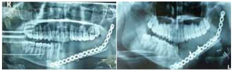
FIG 1 and 2: panoramic radiograph showing adequate bone healing around the implants.
CLINICAL REPORT
A 20-year-old man, who had undergone treatment for ameloblastoma a year ago at the medical hospital, Vellore reported to Dr.Gowd’s dental implant center at Hyderabad, for prosthetic rehabilitation. The medical history revealed that the patient was treated by partial mandibulectomy, and 3 months later 2 Zimmer implants were placed in the fibula bone graft in relation to 33 and 37 respectively.Clinical and radiographic examinations were performed. The mandibular teeth from the left central incisor to the second molar were extracted. The residual dentition was in a good situation with a stable occlusal relationship and was free from periodontal problems. The panoramic radiograph showed that the bone healing process was adequate. However, the height of the grafted bone was limited, and the titanium plates and screws used to hold bone graft remained in place. The graft bone was bucally tilted in the anterior region and lingually tilted in the posterior region. Diagnostic casts and occlusal records were obtained and analyzed. Considering the malposition bone graftand implant placement an implant-supported screw retained hybrid acrylic prosthesis was placed. The soft tissue was thick and easy bleeding. The panoramic radiograph revealed adequate bone healing around the implants. After reshaping the buccal soft tissue by using a soft-tissue laser, we placed custom-made healing caps. Definitive impressions were made using custom trays with additional silicon impression material. The primary interocclusal positions were recorded by using a wax occlusal rim. The corresponding analogs were mounted onto the impression copings. A definitive cast was subsequently poured in type IV dental stone. After impression procedure, it was noticed that, the anterior implant was severely bucally tilted and the posterior implant was lingually tilted with an angle between them being greater than 50 degrees. The casts were transferred and fixed to a semi adjustable articulator. It could be easily noticed that the implants were shifted bucally in the anterior region and lingually in the posterior region. The vertical gap between the top of the implant and the occlusal surfaces was about 15 to 20mm.The cuffs of the implants were as deep as 4 to 8 mm. The screw retained castableabutments were fixed onto the implant analogs, and plastic burnout models were screwed into place. The cantilever was designed to extend medially to restore the occlusal relation of anterior teeth. A wax-up onto the castable abutments were made to connect the implants for a metal framework for the hybrid prosthesis. After the trial placement of the implant abutments and the implant- retained metal framework, an assembly impression was made and the intercuspalocclusal relation was recorded. The master cast was mounted on the articulator. Multiple re-castings were done to get the passive fit. The outer surfaces of the metal framework was colored with a dark pink to give a natural look.
Subsequently, the artificial teeth were arranged. The wax up trial was checked and intra-orally evaluated for retention, esthetics, fit, and proper occlusion. The dentures were fabricated from heat-polymerized acrylic resin. After trial insertion, occlusal adjustments were performed, and the patient was given instructions on denture maintenance and oral hygiene. One month later, the patient was checked again, and recall appointments were subsequently required every 6 months. The patient was satisfied with the treatment and did not present any complications and the panoramic radiograph did not reveal any apparent bone resorption around the implants, though it was noted that on smiling the labially positioned implant in relation to 33 fullness was visible extra orally as a bulge.

FIG 3: Trial placement of the castable implant

FIG 4: A frontal intraoral view of the definitive prosthesis

FIG 5: An occlusal view of the prosthesis

Fig 6: panoramic radiograph did not show apparent bone resorption around the implants after 6 months
DISCUSSION
Ameloblastoma is the most common benign odontogenic tumor, which is characterized by an aggressive infiltration. Wide en bloc resection and subsequent reconstruction with a vascularized bone graft is the optimal treatment.9The vascularized fibula graft was the regular treatment that was used, in the last century, to reconstruct the mandible. Most patients were rehabilitated with removable dentures after segmental resection and suffered functional deterioration. Improper bone dimensions and the massive presence of soft tissue, which may exist in the mandibular reconstruction with other vascularized bone grafts, are still challenging for prosthodontists during the rehabilitation of functional dentition. The patient underwent a vascularized fibula graft after partial mandibulectomy due to ameloblastoma. He was unsatisfied with the traditional dentures because they could not achieve enough retention, stability and support, and they were uncomfortable. Therefore, implant-supported prosthesis became the only available choice. Because of the discrepancy in bone dimensions, the implants were tilted and they were covered by a large amount of soft tissue. The implant-supported bridge was excluded, and animplant screw retained acrylic hybrid prosthesis had to be designed, instead of the ones with individual attachments. Such a design would allow a proper occlusal relationship and, at the same time it is also possible to replace the large defect in both horizontal and vertical plane with a prosthesis which is not very heavy for the load on 2 implants, and achieve adequate loading forces. The occlusal scheme was given at 40 microns under occlusion in centric and all eccentric contacts were relieved thus the tilting and the tipping forces to the implants were minimized and, as a result, this helped prevent bone absorption around the implant. Meanwhile, such a design, which facilitated rehabilitation, provided patients with more functional and esthetic appearance as compared to removable prosthesis. The health of the soft tissues surrounding the implant should also be emphasized. Highly polished custom-made healing caps shaped the deep cuffs of the implants. In terms of self-cleaning, the bottom of the cantilevers were 2 to 3 mm away from the mucosa. All the above would help maintain the soft tissue over the long term.
SUMMARY
The patient described here under-went partial mandibulectomy due to ameloblastoma of the left mandible. After undergoing fibula bone grafting and having 2 implants placed, he was finally rehabilitated with a specially designed implant supported screw retained hybrid acrylic prosthesis. The favorable outcome that we observed suggests that such a design should be considered as an optimal choice when confronted with malpositioned implants and a deep gingival sleeve. Prosthesis supported by the implanted teeth could provide optimized esthetics and functional rehabilitation in patients undergoing mandibular reconstruction. The distribution and loading forces and soft tissue maintenance should be the priorities to consider during the design and the manufacturing of the prosthesis.
REFERENCES
- Goh BT, Lee S, Tideman H, Stoelinga PJ. Mandibular reconstruction in adults: A review. Int J Oral Maxillofac Surg. 2008;37:597–605.
- Functional outcomes of fibula and osteocutaneous forearm free flap reconstruction for segmental mandibular defects. Virgin FW, Iseli TA, Iseli CE, Sunde J, Carroll WR, Magnuson JS, Rosenthal EL. Laryngoscope. 2010 Apr;120(4):663-7. doi: 10.1002/lary.2079
- www.periodonticsltd.com/docs/Gorin_Summer_06.pdf
- Chan MF, Hayter JP, Cawood JI, Howell RA. Oral rehabilitation with implant-retained prostheses following ablative surgery and reconstruction with free flaps. Int J Oral Maxillofac Implants. 1997;12(6):820-7.
- Schepers RH, Slagter AP, Kaanders JH, van den Hoogen FJ, Merkx MA. Effect of postoperative radiotherapy on the functional result of implants placed during ablative surgery for oral cancer. Int J Oral Maxillofac Surg. 2006;35(9):803-8.
- Mericske-Stern R, Zarb GA. Overdentures: An alternative implant methodology for edentulous patients. Int J Prostodont. 1993;6:203-208.
-
Aydin M, Yilmaz A, Katilboglu B, et al. ITI implanta and Dolder bars in the treatment of large traumatic defects of mandible:A clinical report. Dental Traumatol. 2004;20:348-352.
-
.Akca K, Iplikcioglu H, Cehreli MC. A surgical guide for accurate mesiodistal paralleling of implants in the posterior edentulous mandible. J Prosthet Dent. 2002;87:233-235.
-
Mendenhall WM, Werning JW, Fernandes R, et al. Ameloblastoma. Am J Clin Oncol.2007;30:645-648.
Disclaimer from Guident
The views and opinions expressed in articles published in Guident are solely those of the authors and do not necessarily reflect the views of the editors, the editorial board or the publisher, Ivory India. Guident does not endorse any product, technique or treatment described. Clinical content is intended for qualified dental and medical professionals and is not a substitute for individual clinical judgement. Guident, its editors and the publisher accept no liability for any consequence arising from the use of information published in the journal.
How to cite this article
Dr. Vikas Gowd, Dr. Gowds Dental Clinic Hyderabad. Prosthodontic Rehabilitation of Malpositioned Implants After Ameloblastoma Followed by Mandibulectomy and Fibula Bone Graft. Guident. 2018 Mar;11(4). Available from: https://ivoryindia.in/articles/implantology/prosthodontic-rehabilitation-of-malpositioned-implants-after-ameloblastoma-followed-by-mandibulectomy-and.html