Extraction Socket Preservation with Platelet Rich Fibrin Followed by Placement of Implant
Department Of Periodontology The Oxford Dental College, Bangalore, India
ABSTRACT
Tooth extraction being the common dental procedure in the management of tooth decay, fractured root and poor periodontal or endodontic prognosis. Post extraction the socket undergoes a complex process of healing leading to bone resorption and apposition. Replacement of lost teeth gets further complicated for placement of implant if suffïcient volume of bone is not present to achieve primary stability. The preservation of socket thus has become an essential aspect for future therapeutic goal to achieve. Various bone substitutes and autologous biomaterials have been investigated and has proven successful result in preservation of alveolar ridge. This case reports the preservation of extraction socket with placement of platelet rich fibrin followed by placement of implant at 4 months period.
Key word: Extraction socket, Platelet rich fibrin, Delayed implant placement.
Key Message: A case report of a 40 year old male patient was presented where due to poor endodontic prognosis the lower left posterior teeth region had to undergo extraction. The teeth were extracted a traumatically and inoculation of the socket was done. To avoid resorption of the alveolar following the extraction, the sockets were preserved with platelet rich fibrin and hidden X suture was used which enabled to maintain the width of attached gingiva and prevented the collapse of the soft tissue. After 4 months healing period implant placement was done after an adequate amount of bone fill was achieved.
INTRODUCTION
The Implant dentistry aims in restoring the missing teeth to yield aesthetic and functional restorative positions. Following extraction, the socket undergoes a complex process of bone cells migration and maturation leading to selective bone resorption and apposition.11 These further leads to loss of both horizontal and vertical planes of alveolar ridge. This loss of bone volume makes implant placement therapy difficult in later period of time. Therefore adding bone substitutes have shown greater predictability for implant placement for restoration and aesthetic of an individual. Multiple procedures are employed for prevention of post-extraction bone loss and predictable implant placements after extraction, including socket preservation with grafts (biomaterials), and immediate or early implant placements.2 While the clinician has a number of graft materials to choose from, some bone graft materials need longer healing time to achieve even a small amount of new bone incorporation into the graft site (Norton and Wilson, 2002). The autologous biomaterial; Platelet rich fibrin (PRF) is a second-generation platelet concentrate consisting of colossal assemblage ofgrowth factors, which encourages healing and is proposed to be associated with effective and early organization of bone substance and bone volume percentage (Dohan and Choukroun, 2006; Kutkut and Andreana, 2012). The fibrin matrix can be conveniently prepared from autogenous non anti-coagulated blood when centrifuged.3 It is hypothesized that PRF will accelerate socket wound healing after tooth extraction, noticed by increased bone fill and reduced bone resorption. Following the healing of extraction socket implant placement has shown long term stability and success in the rehabilitation of edentulous region. Implant placement in a fully healed alveolar ridge ensures implant insertion and stability.4Therefore the aim of the present case report was to evaluate clinically and radiographically extraction socket healing using autologous platelet rich fibrin (PRF) and membrane and placement of implant at 4 month healing period.
CASE REPORT
A 40 year old male patient came to the department of Periodontics, Bangalore India with a chief complaint of pain in lower posterior teeth region since 2 weeks. The patient gave a history of dull and radiating pain and difficultly in mastication in relation to 35 and 36 region. He had undergone root canal treatment (RCT) in the posterior teeth region (35 36) 2 years back. The intraoral peri apical radiograph revealed radiolucency in mesial root of 36 and 35 and crestal bone loss in relation to 35 and 36 as seen in figure 1. The re RCT was planned but could not be treated due to resorption of the canal and eventually the teeth were planned to be extracted due topoor endodontic prognosis. The patient was explained the procedure for preservation of socket with autologous PRF following the extraction. An informed consent was obtained from the patient before the commencement of the surgical procedure. An inferior alveolar nerve block was given with lidocaine of 1:2,00,000 solution in left posterior teeth region. With the use of periotome the fractured crown and root were removed a traumatically. Following extraction proper inoculation was performed with Gracey curette to remove the peri apical diseased condition.PRF clot and membrane was prepared from patient’s own blood after withdrawing 10 ml of blood from antecubital vein and centrifugingat 2800 rpm for 10 minutes and 2700 rpm for 12 minutes in REMI- 4C centrifugation machine* respectively as depicted in figure 2. The freshly extracted socket was then filled with platelet rich fibrin gel followed byPRF membrane was placed. The sockets were then sutured with hidden X suture5as depicted in figure 3. A cone beam computed tomography (CBCT) was taken at 2 months healing period which illustrated the three dimensional bone density and fabrication of surgical guides enabled the placement of implant in the edentulous ridge as seen in figure.6Before the placement of implant a radiovisiography (RVG) was taken which depictedthe bone fill of the extraction socket at 4 months period as observed in figure 5. A crestal incision was placed in 35 and 36 region. A full thickness mucoperiosteal flap was raised and alveolar bone was visualized. The Genesis implant of 3.3X 13mm dimension was placed after preparation of osteotomy site. Subsequently after achieving the primary stability; abuccal dehiscence was observed. The defect was then further grafted with alloplastic bone graft material. Primary stabilization was obtained by placing horizontal mattress suture followed by placement of periodontal pack. Post-operative RVG was then taken after placement as seen in figure 6.
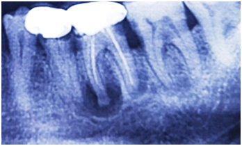
Figure 1: Pre-operative radiographic view in relation to 35 and 36

Figure 2: Preparation of platelet rich fibrin gel and membrane

Figure 3: Socket preservation technique performed with placement of platelet rich fibrin and hidden X suture placed followed by periodontal pack.

Figure 4: Cone beam computed tomography (CBCT) taken at 2months healing period.

Figure 5: Radioviosiography (RVG) taken in relation to 35 and 36 before implant placement at 4 months healing period

Figure 6: Radiovisiography(RVG) taken immediately after implant placement.
DISCUSSION
The present case report mainly highlights the importance of extraction socket healing for implant placement. Following extraction, some amount of internal and external changes takes places which impacts the shape of alveolar ridge. Studies indicate that 100% complete socket fill does not occur. Bone resorption are more pronounced in facial and lingual bone wall mainly in crestal region.7 Preservation of socket further enable final restoration to be placed in aesthetically and functional zone.
The second generation platelet concentrate has been used in various surgical procedures and was successful due to its enhance wound healing process. It wasfurther investigated and confirmed that PRF increases the efficiency of cell proliferation. In addition, alpha platelets in the PRF undergo degranulation (He and Lin, 2009) providing a sustained release of growth factors mainly platelet derived growth factors (PDGF), vascular endothelial growth factor (VEGF), epidermal growth factor (EGF), thrombospondin-1 (TSP-1) and transforming growth factor-beta (TGF-β>)
*- R-4C, REMI Laboratory Instruments, Mumbai, India
which further influencing angiogenesis, epithelialization, stem cell trapping and immune control This PRF are known to also release the growth factors for a longer period of time and its availability at the site of action further optimizes the healing process. It was also known to stimulate osteoblast periodontal ligament cells for regeneration of periodontal defects.8,9 Mazor et al in 2009 stated PRF enhanced new bone formation in sinus augmentation procedures.10Simonpieri et al in a 6 years follow up study elaborated that PRF was sole viable grafting material with long term success in sinus augmentation procedure.11 Thakkar et al in 2016 too found placement of PRF in extraction socket had shown satisfactory result when compared to allograft material. The present case report was in accordance with the study by Dhaval et al in 201612 where preservation of alveolar ridge with adequate amount of bone density was observed at 4th month interval. The maintainenance of width of keratinized tissue both on buccal and lingual wall was due to the hidden X suture which provided an adequate amount of primary closure and preservation of attached gingiva. The present study showed the efficacy of autologous PRF in the healing of extraction sockets. The delayed implant placement allowed proper removal of diseased peri apical lesion was needed and it better remodeling of alveolar ridge. To achieve future osseointegration the implant stability is of adequate importance and mainly depends upon density and dimension of bone surrounding the implant, implant design and surgical technique.13 A poor quality and quantity of bone were considered to be the major risk factor for the implant survival. Miyamoto et al in demonstrated that dental implant stability is positively associated with the thickness of cortical bone thickness.14 The present study too achieved primary stability which was an integral part of osseointegration and was attributed to biomaterial used during ridge preservation procedure. Following achieving the primary stability a buccal dehiscence was observed which was then grafted with alloplastic material without the use of barrier membrane which had shown predictable outcome in maintaining the soft and hard tissues. This was in accordance with study by Cortas et al in 2013 where in 60 delayed implant with buccal dehiscence when treated with bone graft material had achieved a predictable outcome of with minimum soft tissue thickness of 2mm.15
CONCLUSION
From clinical and radio graphical perspective; the use of autologous PRF in the freshly extracted socket enhance new bone fill in 4 month period. Followed by which primary stability was obtained during the implant placement. Further research is warranted in a randomized controlled clinical trial with statistical evaluation along with clinical and radio graphical consideration.
REFERENCES
- Araujo MG, Lindhe J. Dimensional ridge alterations following tooth extraction. An experimental study in the dog. J ClinPeriodontol 2005;32: 212-18.
- Chen ST, Buser D. Ridge preservation techniques for implant therapy. Int J Oral Maxillofac Implants 2009; 24: 260-71.
- Choukroun J, Diss A, SimonpieriA. Platelet-rich fibrin (PRF): a second-generation platelet concentrate. Part V: histologic evaluations of PRF effects on bone allograft maturation in sinus lift. Oral Surg Oral Med Oral Pathol Oral RadiolEndod2006; 101: 299-303.
- Schropp L, Isidor F. Timing of implant placement relative to tooth extraction. J Oral Rehabil2008; 35:33-43.
- Park GJ, Koo KT. The hidden X suture: a technical note on a novel suture technique for alveolar ridge preservation. J Periodontal Implant Sci 2016; 46: 415–25.
- Benavides E, Rios HF, Ganz SD, Resnik R, Reardon GT. Use of cone beam computed tomography in implant dentistry: the International Congress of Oral Implantologists consensus report. Implant Dent 2012;21:78-86.
- Araujo MJ, Lindhe J. Ridge alterations following tooth extraction with and without flap elevation: an experimental study in the dog. Clin Oral Implants Res2009;20:545-49.
- Blair P, Flaumenhaft B. Platelet [alpha]-granules: basic biology and clinical correlates. Blood Rev2009; 23:177-89.
- Kang YH, Jeon SH, Park JY. Platelet-rich fibrin is a Bioscaffold and reservoir of growth factors for tissue regeneration. Tissue Eng Part A2011; 17: 349-59.
- Mazor Z, Horowitz RA, Del Corso M, Prasad HS, Rohrer,DohanEhrenfest DM. Sinus floor augmentation with simultaneous implant placement using Choukroun's platelet-rich fibrin as the sole grafting material: a radiologic and histologic study at 6 months. J Periodontolol2009; 80: 20156-64
- Simonpieri A, Choukroun J, Del Corso M, Sammartino G, Dohan Ehrenfest DM. Simultaneous sinus-lift and implantation using microthreaded implants and leukocyte- and platelet-rich fibrin as sole grafting material: a six-year experience. Implant Dent 2011; 20: 2-12.
- Dhaval J, Deshpande D. A comparative evaluation of extraction socket preservation with demineralized freeze-dried bone allograft alone and along with platelet-rich fibrin: A clinical and radiographic study. ContempClin Dent2016;7: 371–76.
Disclaimer from Guident
The views and opinions expressed in articles published in Guident are solely those of the authors and do not necessarily reflect the views of the editors, the editorial board or the publisher, Ivory India. Guident does not endorse any product, technique or treatment described. Clinical content is intended for qualified dental and medical professionals and is not a substitute for individual clinical judgement. Guident, its editors and the publisher accept no liability for any consequence arising from the use of information published in the journal.
How to cite this article
Dr. Praveen Jayaram, Dr. Koel Debnath, Dr. Anirban Chatterjee. Extraction Socket Preservation with Platelet Rich Fibrin Followed by Placement of Implant. Guident. 2018 Mar;11(4). Available from: https://ivoryindia.in/articles/implantology/extraction-socket-preservation-with-platelet-rich-fibrin-followed-by-placement-of-implant.html