Restoration of Resorbed Mandibular Anterior Ridge Using Implant Supported Prosthesis- a Case Report
Department of Prosthodontics, K.D Dental College & Hospital, Mathura, U.P, India
INTRODUCTION
Clinical management of single missing teeth with dental implants has become common and well accepted. With current techniques and protocols, success of dental implants is more than 90 percent in both the maxilla and mandible, with even higher success in the anterior regions. With the increased popularity of dental implants and a paradigm shift from restoring function to restoring form and function, there has been a greater emphasis on establishing idealized bone foundation and soft tissue contours for optimal esthetic results. Specifically for dental implants in the esthetic zone, intact papillae with ideal facial gingival contours that are indistinguishable from adjacent natural teeth are the goals of therapy and demands of patients.
The goal of hard tissue augmentation is to provide a foundation for ideal implant placement that not only allows for biomechanically sound implant positioning, but also supports soft tissue for optimal esthetics. But one must not over justify the indication for hard tissue augmentation in every case.
Implant placement and restoration to replace single or multiple teeth in the esthetic zone is an especially challenging area for the clinician, particularly in sites with multiple missing teeth and with deficiencies in soft tissue or bone.
CASE REPORT
A 45-year-old male reported with missing mandibular left central incisor and right central & lateral incisor. The patient gave history of trauma due to accident which resulted in loss of left central incisor and right central & lateral incisor. On clinical examination, deficiency in the anterior residual alveolar ridge was observed . On examination there were no clinical signs of periodontitis and dental caries. Clinical and radiographic examination revealed a narrow alveolar ridge.
TREATMENT PROCEDUR
After administration of local anesthesia, an incision was made on the buccal and lingual aspect of the involved edentulous ridge and a full thickness flap was reflected from tooth 31 to tooth 42.
To gain the width of bone, bone shaving was done on the crestal ridge, for the proper placement of implants. Osteotomy was prepared and two tapered implants (BIOHORIZONS) were placed and its stability was verified.
Primary wound closure was obtained by horizontal mattress and interrupted cytoplast 4.0 sutures.
A radiograph was obtained following implant placement. The patient was instructed not to chew or brush in the treated area for at least 4 weeks. Chemical plaque control with chlorhexidine 0.2% (1 minute mouthrinses 3 times a day) was instituted for the same period while Ibuprofen 200 mg was recommended for pain relief.
The patient was seen post surgically after two weeks for suture removal; no untoward sign or symptom was noted. The patient was put on a two week, one month, four month and six month recall ensuring the proper management of implant site.
The patient was then seen after six months since implant placement and the healing abutment was placed for three weeks to achieve soft tissue profile. Then, after three weeks healing abutment was removed and abutment with ball top was placed. A closed tray impression was made with rubber base impression material for the fabrication of prosthesis.
CASE REPORT
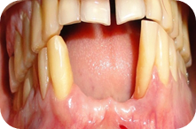
Fig 1: Pre operative
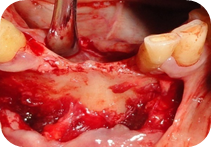
Fig 2: Mucoperiosteal Flap Reflected

Fig 3: Osteotomy Preparation

Fig 4: Surgical Placement of implants

Fig 5: Implant with cover screw
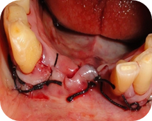
Fig 6: Sutures placed
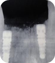
Fig 7: RVG- After surgical placement of implant

Fig 8: Healing abutment placed

Fig 9: Soft tissue profile three weeks after placement of healing abutment

Fig 10: Implant abutment with ball top

Fig 11: Closed tray impression

Fig 12: Angled abutments

Fig 13(a): Final prosthesis - Facial view

Fig 14: Abutment in place.

Fig 15(a): Final Prosthesis in place (Frontal View)

Fig 15(b) : Final Prosthesis in place (Lateral view)
DISCUSSION
For ideal implant placement and optimal esthetic restorations, a comprehensive evaluation of the edentulous site must be performed .
The implant should be considered the apical extension of the restoration and the preferred design of the restoration should guide the surgical placement of the implant. If the desired site is lacking in bone or soft tissue, then augmentation procedures must be employed to create an acceptable site. Optimal esthetic implant restoration depends on proper three-dimensional implant positioning . Four positional parameters contribute to the success of the restoration and all must be carefully considered during implant placement. These are the buccolingual, mesiodistal, and apicocoronal positions relative to the implant platform, as well as the angulation of the implant. Prosthetic design factors (eg, cement- versus screw-retained prosthesis) are also critical.
Regardless of the type of procedure planned for the mature site, proper flap management is critical for success. Careful attention should be paid to incision design and flap extension in an effort to preserve the blood supply of the flap.
REFERENCES
References are available on request.
Disclaimer from Guident
The views and opinions expressed in articles published in Guident are solely those of the authors and do not necessarily reflect the views of the editors, the editorial board or the publisher, Ivory India. Guident does not endorse any product, technique or treatment described. Clinical content is intended for qualified dental and medical professionals and is not a substitute for individual clinical judgement. Guident, its editors and the publisher accept no liability for any consequence arising from the use of information published in the journal.
How to cite this article
Dr. Rahul Nagrath, Dr. Siddharth Sisodia, Dr. Prerna Kaushik. Restoration of Resorbed Mandibular Anterior Ridge Using Implant Supported Prosthesis- a Case Report. Guident. 2014 Jul;7(8). Available from: https://ivoryindia.in/articles/implantology/restoration-of-resorbed-mandibular-anterior-ridge-using-implant-supported-prosthesis-a-case-report.html