Non Surgical Management of Facial Asymmetry with Unilateral Posterior Crossbite in Nongrowing Patient: A Case Report
Department of Orthodontics & Dentofacial Orthopedics, Yenepoya Dental College, Mangalore
ABSTRACT
20-year-old male patient R.B. reported with the complaint of uneven smile and inability to chew. On clinical examination, patient had a straight profile, competent lips, normal nasolabial angle, bilateral class I molar and canine, minimum centric occlusion and centric relation discrepancy, unilateral posterior cross bite extending from 22 to 26, moderate crowding in the upper and lower arches. Maxillary arch was narrow and ‘V’ shaped. Dental midlines were not coinciding. His lower dental midline was shifted to the left by 4 mm.
Treatment included Fixed mechanotherapy, asymmetric unilateral expansion on left side of maxilla and facemask therapy for protraction. Patient was treated with MBT brackets with 0.022 slot and AMEX appliance. When upper arch expanded adequately, lower strap up was carried out.
At the end of treatment, patient had class I molar and canine. Dental midlines were coinciding. Unilateral posterior cross-bite was corrected with good posterior interdigitation. Patient’s facial asymmetry and smile were improved. Currently patient is in retention therapy.
Key words: Unilateral posterior cross-bite, nongrowing patient, transverse malocclusion. AMEX appliance
INTRODUCTION
Posterior crossbite defined as reverse buccal-lingual relation between opposing molars, premolars or both in centric occlusion. The incidence of posterior crossbites ranges from 7% - 23% of the population and the most common form of posterior crossbite is unilateral in nature.1 Unilateral posterior crossbite involves multiple teeth on one side of the occlusion and can be defined either as functional posterior crossbite or as true unilateral posterior crossbite. In functional posterior crossbite, occlusal interferences lead to a lateral shift of the mandible during its closure, to a new position for maximum intercuspation.1-3 True unilateral posterior cross-bite can be distinguished from functional cross-bite by observing the mandibular path of closure and by determining a cross-bite in both centric occlusion (CO) and centric relation (CR) without a functional shift.1-4
The Asymmetric maxillary expansion (AMEX) appliance can be used to treat unilateral posterior crossbites which is fabricated using 0.036-in diameter stainless steel wire consisting of 2 helixes on the crossbite side, and a vertically extending “stopper” on the non-crossbite side.4 The appliance with the stopper was soldered to the molar bands. The appliance was activated by expanding the force arm to a distance equivalent to 8 mm and keeping the arms parallel to each other. This case demonstrated correction of posterior crossbite using AMEX appliance.
PRE-TREATMENT ASSESSMENT
History and clinical examination
R.B. a 20-year- old male patient reported with the complaint of uneven smile and inability to chew. There was no relevant medical or dental history.
Extraoral examination revealed he had a straight profile, competent lips, normodivergent, normal mentolabial sulcus, facial asymmetry, and mandible deviated to left side (Figure 1).
Intra oral examination revealed patient had permanent dentition in upper and lower arches up to Second molars in the oral cavity. He presented with normal gingiva, frenal attachment with narrow upper arch with moderate crowding. Patient had heavily restored 26 and dental Caries 36, 46. Right buccal segment showed Class I molar and canine relations and left buccal segment showed Class I molar and canine (in crossbite) (Figures 2 and 3), minimum CO and CR discrepancy, unilateral posterior cross bite extending from 22 to 26, moderate crowding in the upper and lower arches. Maxillary arch was narrow and ‘V’ shaped. Dental midlines were not coinciding and his lower dental midline was shifted to the left by 4 mm (Figures 1 and 2).
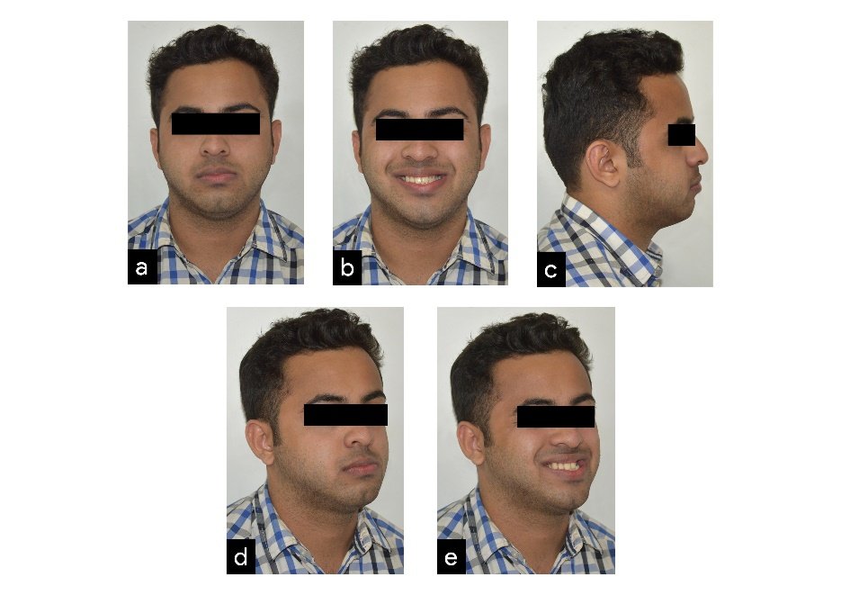
Figure 1. Pre-treatment extraoral photographs (a) Frontal, (b) frontal smiling, and (c) profile (d) oblique, (e) Oblique Smiling

Figure 2. Pre-treatment intraoral photographs- (a) Right buccal (b) frontal, (c) left buccal, (d) upper occlusal, and (e) lower occlusal

Figure 3. Pre-treatment Study Model (a) Right buccal (b) frontal, (c) left buccal, (d) upper occlusal, and (e) lower occlusal
Pre-treatment radiographs taken were lateral cephalogram and Orthopantomogram (OPG). (Figure 4) and cephalometric analysis showed skeletal class I relation, with deficient maxilla as compared to mandible, normally aligned upper and lower anteriors, and vertical growth pattern
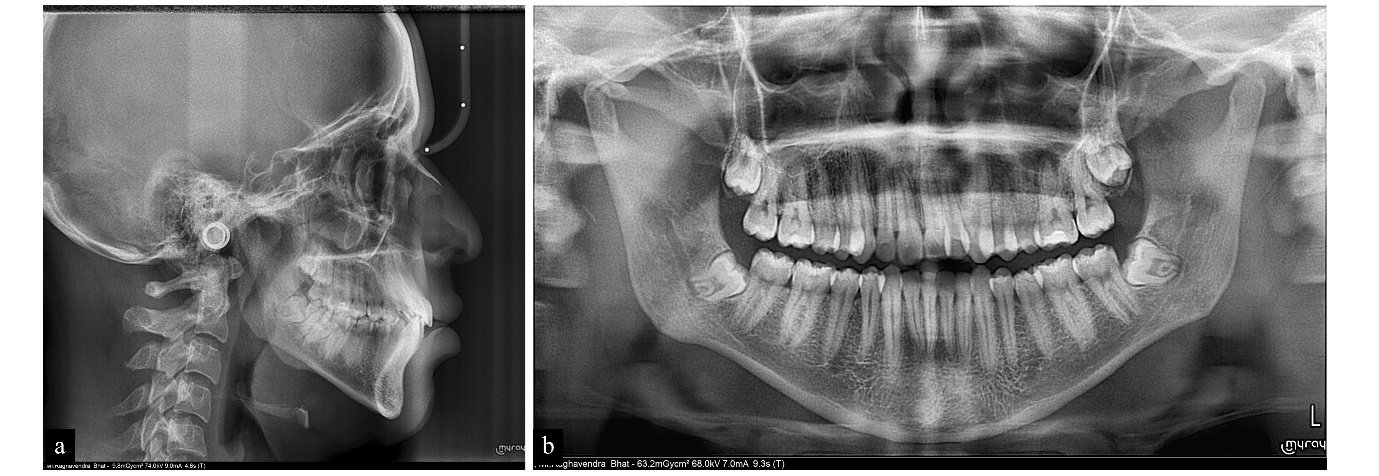
Figure 4. Pre-treament radiographs- (a). Lateral Cephalogram, (b). Panoromic radiograph
Model Analysis
Bolton’s analysis showed overall mandibular tooth material excess by 3.05 mm and anterior mandibular tooth material excess of 1.7 mm
Diagnostic summary
Angle’s Class I with Dewey’s type 4 malocclusion (Class I molar relation with posterior cross-bite), vertical growth pattern, and moderate crowding in anterior region and non-coinciding dental midlines.
Problems List
- Unilateral posterior cross-bite
- ‘V’ shaped upper arch
- Upper Arch Constriction
- Moderate crowding in upper and lower arches
- Dental midlines not coinciding
- Uneven smile
- Rotated 35,44, and 45
- Facial asymmetry
Treatment objectives
The following treatment objectives were established
- Correction of unilateral posterior cross bite
- Correction of midline
- Correction of crowding
- Protraction of maxilla
- Restriction of mandible
- To improve facial symmetry
- To improve smile
- To improve posterior interdigitations
- To establish ideal overjet and overbite
- To achieve balanced soft tissue profile

Figure 5. Upper strap-up a) Right buccal (b) frontal, (c) left buccal, (d) upper occlusal, and (e) lower occlusal

Figure 6. Facemask therapy: a. Frontal, b. Oblique, c. Profile

Figure 7. Lower strap-up a) Right buccal (b) frontal, (c) left buccal, (d) upper occlusal, and (e) lower occlusal
Treatment Plan
The treatment plan suggested was fixed mechanotherapy with facemask and unilateral expansion using AMEX to expand the maxilla on the left side. Facemask was used to protract the maxilla
Proposed retention strategy
In the lower arch, fixed retainer is planned. In the upper arch, retainer incorporated with expansion screws would be delivered to reactivate if required. His all third molars which are impacted would be advised for extraction after orthodontic treatment.
Treatment progress
The patient was treated with a preadjusted edgewise appliance, MBT prescription (Victory series, 3M Unitek, slot dimensions 0.022″ × 0.028″). Brackets were initially bonded to the maxillary arch and 0.014 round NiTi arch wire was placed in the maxillary arch for the initial levelling and aligning phase [Figure 5]. AMEX was fabricated, cemented to molars [Figure 5] and activated in patient’s mouth. Facemask therapy was started along with orthodontic strap up to protract the maxilla [Figure 6]. The 0.014 round NiTi AW were then followed by 0.016 round NiTi AW, 0.016 × 0.022 NiTi. Once upper arch was aligned, the lower arch was bonded and 0.014 round NiTi wire was placed in the mandibular arch [Figure 7]. The 0.016 × 0.022 NiTi wires were then followed by 0.019 × 0.025 NiTi wires. Then 0.019 × 0.025 SS wires were placed in the upper and the lower arch. Patient was recalled at monthly interval to monitor the progress. Elastics were given to settle the occlusion before debanding (Figures 8). After 18 months of commencement of treatment, brackets were removed in upper arch followed the lower arch (Figures 9 and 10).
Post treatment Assessment
The post treatment results showed that all objectives were achieved (Figures 10 to 12). Unilateral posterior cross-bite was corrected with good posterior interdigitations. Root parallelism were seen except for upper left lateral incisor in post treatment OPG (Figures 13). His smile aesthetics has greatly improved and chin prominence was reduced. Post treatment cephalometric assessment showed that incisors are well aligned, nasolabial angle improved, lower lips are normally placed, and maxillary length increased (Table 1). Superimposition showed maxillary arch expanded considerably (Figure 14).

Figure 8. Settling Photographs. (a) Right buccal (b) frontal, (c) Left buccal

Figure 9. Post upper debanding photographs. (a) Right buccal (b) frontal, (c) left buccal, (d) upper occlusal, and (e) lower occlusal

Figure 10. Post treatment Intraoral Photographs. (a) Right buccal (b) frontal, (c) left buccal, (d) upper occlusal, and (e) lower occlusal (f) Retainer with expansion screws

Figure 11. Post treatment extraoral Photographs. (a) Frontal, (b) frontal smiling, and (c) profile (d) oblique, (e) Oblique Smiling

Figure 12. Post treatment study model. (a) Right buccal, (b) frontal, (c) left buccal, (d) upper occlusal, and (e) lower occlusal
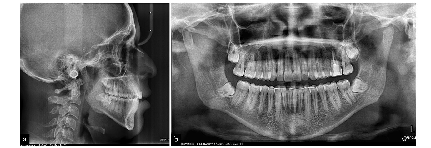
Figure 13 Post-treament radiographs- (a). Lateral Cephalogram, (b). Panoromic radiograph

Figure 14. Pre and Posttreatment Superimposition, (a) Overall, (b) maxilla, and (c) mandible, (d). maxillary cast superimposition
DISCUSSION
Careful assessment of the characteristics of unilateral posterior crossbites is required for successful treatment outcome. Early orthodontic intervention is desirable to treat unilateral posterior crossbites. If left untreated, it may require a combination of orthodontics and surgery. Orthognathic surgery has the disadvantages of surgical risk and cost. In view of risk-benefit analysis, if correcting skeletal Class I asymmetry is the only goal, the patient's satisfaction level may be low even after the surgery. So, a non-invasive management of unilateral posterior crossbites was decided in this patient.
Non-invasive treatment of unilateral posterior crossbite involves expansion of the maxillary arch, removal of occlusal interferences and elimination of the functional shift.3,5-7 W-arches and quad helix appliances can be modified by changing the length of the arms to include more teeth in the anchorage unit.3 A modified quad-helix appliance has been designed to produce asymmetrical expansion in patient with unilateral cleft lip and palate patient.8 Studies have shown that AMEX appliance also proved to be effective for treating unilateral posterior crossbites.4 Since the ratio of maxillary and mandibular length were disproportionate (according to Mc Namara norms),9 we decided to give facemask in addition to AMEX by making use of synergistic effects for protraction along with expansion.
At the end of eighteen months of active treatment, facial asymmetry was corrected, chin prominence reduced, Smile aesthetic improved. Transverse discrepancy was corrected with good interdigitation of posterior teeth

Table 1. Pre treatment and Post treatment Cephalometric Values
CONCLUSIONS
This case report demonstrates the efficiency of the combined clinical use of AMEX and Facemask with fixed appliance in growing patient to treat unilateral posterior cross-bite effectively.
REFERENCES
- Kennedy DB, Osepchook M. Unilateral posterior crossbite with mandibular shift: a review. J CanDent Assoc. 2005;71(8):569-73.
- Proffit WR, Fields HW, Sarver DM, Ackerman JL. 2013. Contemporary Orthodontics. 5 ed. 2013: Mosby Publication; St. Louis, Missouri.
- Hesse KL, Artun J, Jondeph DR, Kennedy DB. Changes in condylar position and occlusion associated with maxillary expansion for correction of functional unilateral posterior crossbite. Am J Orthod Dentofacial Orthop 1997;111(4):410–8.
- Toroglu MS, Uzel E, Kayalioglu M, Uzel I. Asymmetric maxillary expansion (AMEX) appliance for treatment of true unilateral posterior crossbite. Am J Orthod Dentofacial Orthop. 2002 Aug;122(2):164-73.
- Bjerklin K. Follow-up controls of patients with unilateral posterior cross-bites treated with plates or the quad-helix appliances. Am J Orofac Orthop 2000; 61(2):112–24.
- Ranta R. Treatment of unilateral posterior crossbite: comparison of the quadhelix and removable plate. ASDC J Dent Child 1988; 55(2):102–4.
- Hermanson H, Kurol J, Ronnerman A. Treatment of unilateral posterior crossbite with quad-helix and removable plate. Europ J Orthod 1985; 7(2):97–102.
- Enacar A,O¨ zgen M. Asymmetric maxillary expansion appliance (ABHE). Cleft Palate Craniofac J 1993;30:416-7.
- McNamara JA. A method of cephalometric evaluation. Am J Orthod. 1984;86:449-69.
Disclaimer from Guident
The views and opinions expressed in articles published in Guident are solely those of the authors and do not necessarily reflect the views of the editors, the editorial board or the publisher, Ivory India. Guident does not endorse any product, technique or treatment described. Clinical content is intended for qualified dental and medical professionals and is not a substitute for individual clinical judgement. Guident, its editors and the publisher accept no liability for any consequence arising from the use of information published in the journal.
How to cite this article
Dr. Shahistha Parveen D, Dr. Rohan Mascarenhas. Non Surgical Management of Facial Asymmetry with Unilateral Posterior Crossbite in Nongrowing Patient: A Case Report. Guident. 2021 Sep;14(10). Available from: https://ivoryindia.in/articles/orthodontics/non-surgical-management-of-facial-asymmetry-with-unilateral-posterior-crossbite-in-nongrowing-patient-a-case.html